

Journal of Veterinary Science & Medicine
Download PDF
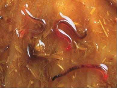 Figure 1:Adult large strongyles. Source: Urquhart et al.
[9].
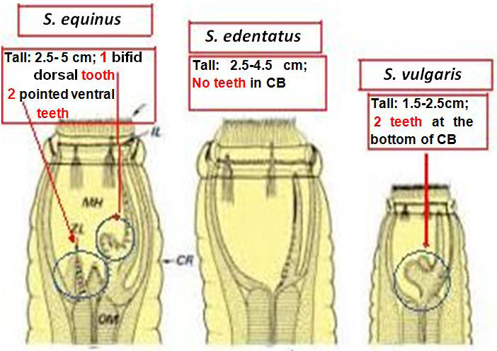 Figure 2:Identification of adult Strongyle species in equines. Source:
Urquhart et al. [9].
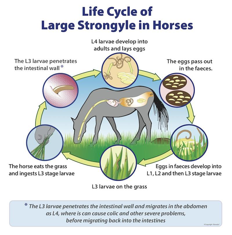 Figure 3:Life cycle of large strongyle parasites. Source: Saeed et al.
[14].
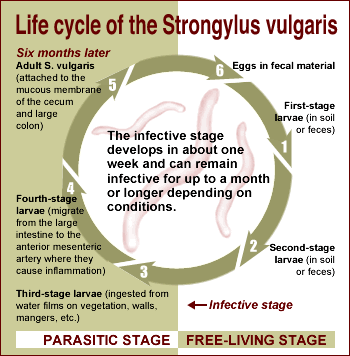 Figure 4:Life cycle of Strongylus vulgaris. Source: Saeed et al. [14].
The epidemiology of equine strongylosis is influenced by environmental conditions, host age, season, and management practices. Seasonal variation in infection is closely associated with temperature and moisture, which affect the development, survival, and availability of free-living larval stages on pastures. In regions characterized by seasonal rainfall, favorable temperatures and high relative humidity may promote larval development and increase the risk of infection during wet periods [17]. In areas with cold winters and mild summers, strongyle egg shedding and pasture contamination may increase during spring and remain high throughout summer, when environmental conditions favor the development of infective larvae. These larvae may persist into early autumn, increasing exposure among susceptible horses. Conversely, hot and dry summer conditions can restrict larval development and survival; however, continued egg shedding and reinfection may sustain pasture contamination [17].
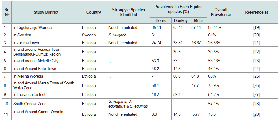 Table 1:Worldwide prevalence of Equine Strongyles within the recent 10 years.
Host age is an important determinant of strongyle infection intensity and egg shedding. Foals, yearlings, and young horses, particularly those less than four to five years old, may carry heavier parasite burdens and shed more eggs per gram of feces than older animals. In contrast, adult horses generally develop partial immunity that helps regulate parasite burdens and egg production. However, this immunity is incomplete, and adult horses may continue to shed eggs, thereby contributing to pasture contamination [5,30]. Factors that compromise host condition, including inadequate nutrition, excessive workload, and other sources of physiological stress, may further increase susceptibility to infection or aggravate its consequences [5]. Although age can influence parasite burden and egg shedding, the reported relationship between sex and fecal egg counts should be interpreted in the context of the population and study conditions, rather than assumed to be universal [30].
Management practices also play a major role in determining exposure to infective larvae and the persistence of pasture contamination. Overcrowding, poor sanitation, inadequate pasture management, and the accumulation of feces in grazing areas can increase the likelihood of infection. Strongylosis may therefore affect adult horses maintained in suburban paddocks when stocking density is high and management is inadequate [9]. Additional factors influencing infection intensity and disease severity include nutritional deficiencies, the availability and quality of feed, host immune status, grazing behavior, seasonal variation, and the number of infective L3 larvae ingested. These factors may interact to influence parasite establishment, persistence, and clinical outcomes. Consequently, differences in husbandry, stocking density, pasture hygiene, and parasite-control practices can contribute to variation in strongyle infection among equine populations and production systems [9].
The principal pathogenic effects of S. vulgaris arise from the vascular lesions caused by its migrating larvae. As the larvae migrate through the cranial mesenteric artery and its branches, they induce endarteritis and thrombus formation, which may compromise blood supply to the intestine. The resulting ischemia can cause intestinal infarction and, in severe cases, necrosis of the intestinal wall. Associated complications may include intestinal dysfunction, ileus, and, occasionally, intestinal rupture. Thromboemboli detached from the primary lesions may obstruct smaller arteries, further compromising tissue perfusion. Verminous aneurysms involving the cranial mesenteric artery and its branches may also contribute to colic, partly through associated vascular inflammation and irritation of adjacent nerve plexuses. Clinical manifestations depend on the severity and location of the vascular lesions and may range from intermittent abdominal pain to severe, potentially fatal colic [9,31].
In addition to vascular injury, large strongyles can cause tissue damage during larval migration and adult establishment in the large intestine. The larvae of S. equinus may produce hemorrhagic tracts in the liver and pancreas, whereas those of S. edentatus can cause inflammatory lesions and hemorrhagic nodules in tissues along their migratory pathways, particularly the liver, peritoneal tissues, and intestinal wall [8]. The emergence of developing parasites into the intestinal lumen may also disrupt the intestinal mucosa and contribute to local inflammation [8].
The severity of clinical manifestations depends on several factors, including the parasite species, infection intensity, host age, nutritional status, immune response, and previous exposure to infection. Large strongyle adults possess well-developed buccal capsules that facilitate attachment to the intestinal mucosa and feeding, potentially causing mucosal injury and blood loss. Consequently, heavy infections may contribute to anemia, weight loss, and reduced performance [16]. The clinical effects of migrating larvae may become apparent during the weeks or months following infection, depending on the species and its developmental pathway. In some horses, particularly those with previous exposure, substantial pathological lesions may develop without obvious clinical signs. Nevertheless, severe larval migration and associated tissue damage can result in significant disease [16].
The principal lesions associated with migrating S. vulgaris larvae occur in the cranial mesenteric artery and its major branches. Larval migration damages the vascular endothelium, inducing endarteritis, thickening of the arterial wall, and thrombus formation. These changes may compromise intestinal blood supply and result in ischemia, infarction, and necrosis of affected intestinal tissues. In some cases, true aneurysms, characterized by abnormal arterial dilatation and thinning of the vessel wall, may develop. Such lesions are commonly associated with the term “verminous aneurysm,” although aneurysm formation is not an inevitable consequence of infection [34,31].
In addition to vascular injury, other large strongyle species produce characteristic lesions during larval migration. Migrating larvae of S. equinus and S. edentatus may cause hemorrhagic tracts in the liver and other tissues along their migratory pathways. At postmortem examination, hepatic lesions may include hemorrhages, fibrous scarring beneath the hepatic capsule, and inflammatory changes. Sub peritoneal hemorrhage, hematoma formation, peritonitis, and omental adhesions may also occur. In the intestinal wall, migrating larvae may induce nodules and hemorrhagic foci, while larval migration through vascular tissues may result in perivascular inflammation and thickening [31,8].
The overall pathogenic significance of strongyle infection lies in its capacity to cause tissue injury, impair intestinal function, compromise blood supply, and reduce the general condition and productivity of affected equids. The severity of these effects depends on the parasite species, infection burden, nutritional status, and physiological condition of the host. Heavy infections and severe pathological complications may be fatal, particularly when extensive vascular damage or intestinal injury occurs [8,31].
Fecal examination is one of the most widely used diagnostic approaches. Fecal flotation can detect strongyle-type eggs, while the number of eggs per gram of feces (EPG) can be estimated using quantitative methods to assess egg-shedding intensity [16,34]. However, fecal egg counts primarily indicate the presence and eggshedding activity of patent adult infections; they do not reliably measure the total parasite burden or the severity of disease caused by migrating larvae. Furthermore, strongyle eggs are morphologically similar, and routine fecal microscopy generally cannot differentiate large strongyles from cyathostomins or identify individual species, particularly in mixed infections [19].
A negative fecal examination does not exclude strongyle infection, especially during the prepatent period, when immature parasites have not yet begun producing eggs. The original source states that eggs may be absent in animals younger than 9–12 months because of the prolonged prepatent period of large strongyles; however, this age-based statement should be interpreted cautiously because the prepatent period varies among species, and age alone cannot determine whether an infection is patent [8].
Fecal culture, or coproculture, is useful for differentiating strongyle groups when combined with morphological examination of the resulting third-stage larvae (L3). This method involves incubating fecal samples under suitable conditions to allow eggs to hatch and larvae to develop before the larvae are recovered and identified. Although coproculture can help distinguish large strongyles from cyathostomins, species-level identification may be difficult because larval morphology can overlap among taxa [16].
Pasture management measures include regular removal of feces from paddocks, appropriate pasture rotation, avoidance of excessive stocking density, and careful management of grazing areas. Where feasible, mixed-species grazing may help reduce the accumulation of host-specific parasites, although its effectiveness depends on the animal species involved and the local management system. Grazing different age groups separately may also help manage exposure, particularly where young horses are more susceptible to heavy infection. However, pasture rotation alone may not eliminate infective larvae because their survival varies with environmental conditions [7,9]].
Anthelmintic treatment should be based on parasite burden, epidemiological risk, clinical condition, and evidence of drug efficacy rather than relying exclusively on fixed treatment intervals. Although routine treatment of all horses at regular intervals has historically been recommended, indiscriminate and frequent deworming can increase selection pressure for anthelmintic resistance. Modern control programs therefore emphasize veterinary-guided treatment, fecal egg count monitoring, and fecal egg count reduction testing to assess treatment effectiveness and help identify suspected resistance.
Treatment decisions should also account for the fact that fecal egg counts do not reliably detect encysted cyathostomin larvae or migrating stages of large strongyles [7,9,16].
1. Equine owners should minimize excessive workloads and provide adequate nutrition, rest, and appropriate husbandry to maintain animal health and resilience.
2. Equine owners and caretakers should receive education on strongyle transmission, clinical signs, prevention, and appropriate parasite-management practices.
3. Strategic, veterinarian-guided anthelmintic treatment and improved pasture management should be implemented to reduce parasite burdens and minimize pasture contamination.
4. Newly introduced equines should be managed separately from resident animals until their parasite status has been assessed. Quarantine procedures, fecal examination, and veterinarian-guided treatment should be implemented as appropriate, followed by reassessment when indicated. A fixed isolation period of 48–72 hours alone does not guarantee that newly introduced animals are free of parasites.
5. Housing and grazing management should reduce overcrowding and, where feasible, limit the accumulation and transmission of infective larvae through regular manure removal and appropriate pasture management. 6. Governmental and veterinary authorities should support equine health-promotion programs that improve access to veterinary services, owner education, diagnostic facilities, and sustainable parasite-control strategies.
7. Anthelmintic efficacy should be monitored where feasible, and treatment decisions should be guided by veterinary advice and fecal egg count results to help detect and manage anthelmintic resistance.
Review Article
Review of Equine Strongyle Parasites: Biology, Ecology, And Pathogenic Importance
Mequannet Fentie¹, Tewodros Alemneh¹*, and Mekonen Baylie²
¹Woreta Town Agriculture, and Livestock and Fishery Resources
Development Office, South Gondar Zone, Woreta, Ethiopia
²Fogera District Livestock and Fishery Resources Development Office, South Gondar Zone, Woreta, Ethiopia
²Fogera District Livestock and Fishery Resources Development Office, South Gondar Zone, Woreta, Ethiopia
*Address for correspondence:Tewodros Alemneh, Department of Veterinary, Woreta Town Agriculture, and Livestock and Fishery Resources Development Office, South
Gondar Zone, Woreta, Ethiopia. E-mail Id: tedyshow@gmail.com
Submission: 03 August, 2026
Accepted: 10 November, 2026
Published: 16 November, 2026
Copyright: © 2026 Fentie M, et al. This is an open access article
distributed under the Creative Commons Attribution License, which
permits unrestricted use, distribution, and reproduction in any medium,
provided the original work is properly cited.
Keywords:Strongyles; Equines; Biology; Ecology; Pathogenicity
Abstract
Equine strongylosis, caused by large and small strongyles, is one of the most
important gastrointestinal parasitic infections affecting equines worldwide. These
nematodes, belonging to the family Strongylidae, have complex life cycles, and
some species exhibit larval migration through host tissues, causing damage to
multiple organs. However, their pathogenicity and the severity of associated disease
vary among species. Large strongyles, including Strongylus vulgaris (S. vulgaris), S.
edentatus, and S. equinus, are particularly important because of their potential to
cause severe disease. Their major pathogenic effects include verminous arteritis,
vascular obstruction, thrombosis, embolic complications, and visceral tissue damage,
particularly during larval migration; severe infections may be fatal. In contrast, small
strongyles (cyathostomins) are highly prevalent in many equine populations and can
cause clinical signs such as diarrhea, reduced appetite, weight loss, poor coat condition,
and lethargy. Severe cyathostomin infections, particularly those associated with the
emergence of encysted larvae, may cause substantial intestinal damage. Despite
advances in diagnostic techniques and the availability of anthelmintic treatments,
strongyle infections remain important concerns for equine health, welfare, and
productivity. Effective control requires an integrated approach combining appropriate
anthelmintic treatment, parasitological monitoring, and sound pasture management.
These measures can reduce parasite burdens and pasture contamination while
supporting the responsible use of anthelmintics and helping to limit the development
of anthelmintic resistance. Therefore, this review aimed to provide an overview of the
biology, ecology, pathogenicity, diagnosis, treatment, and control of equine strongyles,
emphasizing their implications for equine health and welfare.
Introduction
Equine strongylosis, a prevalent disease among grazing equids,
results from infection by a group of nematode parasites collectively
known as strongyles. The disease occurs when horses graze on
pastures contaminated with strongyle larvae that hatch from eggs
shed in the feces of infected animals. Equines provide substantial
economic benefits in Ethiopia, particularly for transportation and
agriculture [1]. The majority of equine populations are concentrated
in areas of high human population density, where crop production
and livestock rearing are the dominant agricultural activities [2].
The term “strongyle” (commonly referred to as a redworm) denotes nematodes belonging to the family Strongylidae. This family comprises two key subfamilies: Strongylinae (large strongyles) and Cyathostominae (small strongyles, cyathostomins, or small redworms). In equines, strongylosis caused by large and small strongyles is among the most economically important nematode infections. Large strongyles are generally recognized as important internal parasites of horses, with the major pathogenic species being Strongylus vulgaris (S. vulgaris), S. edentatus, and S. equinus. Their adult stages reside in the large intestine, particularly the caecum and colon. Based on their mouthparts, S. vulgaris is known as the doubletoothed strongyle, S. edentatus as the toothless strongyle, and S. equinus as the triple-toothed strongyle. Among these three species, S. vulgaris is smaller than the other two large strongyle species [3].
The term “strongyle” (commonly referred to as a redworm) denotes nematodes belonging to the family Strongylidae. This family comprises two key subfamilies: Strongylinae (large strongyles) and Cyathostominae (small strongyles, cyathostomins, or small redworms). In equines, strongylosis caused by large and small strongyles is among the most economically important nematode infections. Large strongyles are generally recognized as important internal parasites of horses, with the major pathogenic species being Strongylus vulgaris (S. vulgaris), S. edentatus, and S. equinus. Their adult stages reside in the large intestine, particularly the caecum and colon. Based on their mouthparts, S. vulgaris is known as the doubletoothed strongyle, S. edentatus as the toothless strongyle, and S. equinus as the triple-toothed strongyle. Among these three species, S. vulgaris is smaller than the other two large strongyle species [3].
Equine strongylosis is one of the most common parasitic infections
of horses worldwide, with substantial economic consequences
and adverse effects on animal welfare. It occurs wherever equine
populations are present [4]. In Ethiopia, S. vulgaris has been reported
to be common among equids, particularly in mid- and high-altitude
areas characterized by relatively high rainfall and a bimodal rainfall
pattern. Among the gastrointestinal nematodes of equines, large
strongyles, particularly S. vulgaris, have long been considered
important pathogens because of their capacity to cause serious disease
[5]. Although S. vulgaris was once regarded as the most important
parasitic threat to horses, its significance has declined following the
introduction and use of modern anthelmintic compounds [6].
The pathogenicity of large strongyles is closely associated with
the migration of their larval stages through different tissues and
organs. When equines ingest infective larvae, the parasites develop
and migrate through the host before reaching the large intestine,
where the adults establish themselves. Strongylus vulgaris migrates
to the anterior mesenteric artery, S. edentatus to the liver and flank
region, and S. equinus to the liver and pancreas. These distinct
migration patterns contribute to the pathological effects associated
with infection by the different species [6].
Diagnosis of equine strongylosis involves several methods, including fecal examination for strongyle egg detection, fecal culture for larval identification, and clinical or postmortem assessment of associated pathological changes. In particular, per rectal examination may reveal abnormalities associated with aneurysm of the cranial mesenteric artery in cases involving S. vulgaris [3]. Equines are treated with anthelmintic drugs to eliminate adult strongyles from the large intestine and prevent excessive pasture contamination with eggs and infective third-stage larvae (L3). Effective control programs therefore combine strategic anthelmintic treatment with improved pasture management to reduce parasite transmission and pasture contamination [7].
Diagnosis of equine strongylosis involves several methods, including fecal examination for strongyle egg detection, fecal culture for larval identification, and clinical or postmortem assessment of associated pathological changes. In particular, per rectal examination may reveal abnormalities associated with aneurysm of the cranial mesenteric artery in cases involving S. vulgaris [3]. Equines are treated with anthelmintic drugs to eliminate adult strongyles from the large intestine and prevent excessive pasture contamination with eggs and infective third-stage larvae (L3). Effective control programs therefore combine strategic anthelmintic treatment with improved pasture management to reduce parasite transmission and pasture contamination [7].
Therefore, this review aimed to examine the biology, ecology,
pathogenic significance, clinical signs, diagnosis, treatment, and
control strategies of equine strongylosis; raise awareness among
stakeholders about its economic impact; and enhance understanding
of the effects of strongyle infections on equine health and productivity.
Biology of Strongyles
Taxonomic and Morphological Classification:
Large strongyles are classified under the kingdom Animalia,
phylum Nemathelminthes, class Nematoda, order Strongylida,
superfamily Strongyloidea, family Strongylidae, and subfamily
Strongylinae [8]. They are dioecious, with distinct male and female
worms, and females are oviparous, producing strongyle-type eggs
through sexual reproduction. Like other nematodes, they lack
specialized circulatory and respiratory systems. Grossly, these
parasites appear as robust, dark red worms [Figure 1] that are readily
visible against the intestinal mucosa. They possess a cylindrical,
unsegmented body, a body cavity, an intestine, an anus, and a
nervous system. Some species also possess a prominent leaf crown
surrounding the mouth opening [9].
Morphological differentiation of strongyle species in horses is
primarily based on characteristics of the cephalic end (head) of the
worm [Figure 1]. The buccal capsule is well developed and exhibits
structural features that aid species identification. In many strongyles,
a distinct dorsal thickening within the buccal capsule, known as
the dorsal gutter, is present. The mouth collar, a cuticular structure
surrounding the mouth opening, may bear submedian and lateral
cephalic papillae. Leaf-like structures called leaf crowns occur along
the anterior margin of the mouth collar. Typically, two leaf crowns
are present: the external and internal crowns, with the internal crown
located distal to the external crown. In some genera, the external leaf
crown is supported by an additional sclerotized ring, referred to as
the extra-chitinous support, located anterior to the buccal capsule [9]
Adult large strongyles possess a well-developed buccal capsule,
while males have a prominent copulatory bursa. Their characteristic
reddish coloration gives rise to the common name “redworms.”
Species differentiation relies primarily on body size and the presence,
absence, number, and shape of teeth within the buccal capsule.
Strongylus vulgaris (S. vulgaris) measures 1.5–2.5 cm in length and
possesses two ear-shaped, rounded teeth. Strongylus edentatus (S.
edentatus) measures 2.5–4.5 cm and lacks teeth, whereas Strongylus
equinus (S. equinus) measures 2.5–5 cm and possesses three conical
teeth, one of which is dorsally positioned and larger than the other two
[Figure 2]. The shape of the buccal capsule also differs among these
species and can assist in morphological identification
[9] [Figure 2].
Life Cycle:
Equine strongyles have a direct life cycle, with transmission
occurring through the ingestion of infective third-stage larvae
(L3) from contaminated herbage. Although large strongyles and
cyathostomins share a similar general life cycle, they differ in their
larval migration, development, and pathogenic effects within the host.
The larvae of large strongyles migrate through different tissues and
organs before returning to the large intestine, where they mature into
adults. Strongylus vulgaris larvae migrate to the cranial mesenteric
artery and its branches, whereas S. edentatus and S. equinus larvae
migrate through other tissues, including the liver, pancreas, and
perirenal and retroperitoneal tissues [10]. These differences in
migration patterns contribute to species-specific pathological effects
and variations in the prepatent period. In large strongyles, the
prepatent period—the interval between ingestion of infective larvae
and the first detection of eggs in feces—generally ranges from 6 to 12
months, depending on the species
[11,9].
The free-living stages of the life cycle begin when eggs produced
by adult female strongyles are shed in the feces into the environment.
Under suitable temperature and moisture conditions, the eggs hatch
and release first-stage larvae (L1), which develop into second-stage
larvae (L2) and subsequently molt into infective third-stage larvae (L3).
Development is influenced by environmental conditions, including
temperature and moisture; under favorable summer conditions,
L3 may develop within approximately 10 days to 2 weeks after the
eggs are passed. The L3 retains an outer protective sheath, which
increases its resistance to environmental stress, including chilling
and dehydration, compared with the earlier larval stages. Adequate
moisture facilitates larval movement from feces onto surrounding
herbage, whereas dehydration can restrict this movement and reduce
the likelihood of transmission. Larvae generally remain relatively close
to the soil surface, although their vertical and horizontal distribution
depends on environmental conditions. Rainfall can facilitate the
movement of infective larvae from fecal deposits onto herbage,
thereby increasing the likelihood of ingestion by grazing equids. The
five developmental stages recognized in the strongyle life cycle are L1,
L2, L3, fourth-stage larvae (L4), and fifth-stage larvae (L5), followed
by the adult stage (Figure 3). Infection occurs when equids ingest
L3-contaminated herbage, after which larval development proceeds
within the host [2].
Following ingestion, the developmental pathways of large
strongyles and cyathostomins diverge. Large strongyle larvae undergo
extensive tissue migration before returning to the large intestine to
mature. Although the usual prepatent period of large strongyles is
approximately 6–12 months, the duration varies with species and
developmental conditions [9]. Occasionally, migrating larvae may
enter aberrant locations, including the kidneys, thoracic cavity,
or testes [12]. In contrast, small strongyle larvae (cyathostomins)
penetrate the mucosa and submucosa of the caecum and colon, where
they may become encysted and undergo arrested development. These
encysted larvae can persist for extended periods, reportedly up to
approximately 2.5 years, before resuming development and emerging
into the intestinal lumen [13]. Thus, differences in tissue migration
and larval persistence distinguish the biology of large strongyles from
that of cyathostomins and contribute to their differing patterns of
pathogenicity [8,13].
Life cycle of S. vulgaris:
Following ingestion, the infective third-stage larvae (L3) penetrate
the intestinal mucosa and molt into fourth-stage larvae (L4) within
the submucosa. The larvae then enter small arteries and migrate along
the vascular endothelium toward their predilection site in the cranial
mesenteric artery and its major branches. After several months of
development and migration, the larvae molt into fifth-stage larvae
(L5) and eventually return toward the intestinal wall through the
arterial circulation. Nodules develop around the larvae, primarily
in the walls of the caecum and colon. As the larvae mature, these
nodules rupture, releasing young adult parasites into the intestinal
lumen, where they develop into sexually mature adults. The prepatent
period is approximately 6–7 months [2] [Figure 4].
Life cycle of S. edentatus:
After penetrating the intestinal mucosa, the infective third-stage
larvae (L3) enter the portal circulation and reach the liver parenchyma
within a few days. Approximately two weeks after infection, they
molt into fourth-stage larvae (L4), and migration continues within
the liver. By 6–8 weeks post-infection, larvae can be found beneath
the peritoneum, particularly around the hepatorenal ligament.
They subsequently migrate subperitoneally to various sites, with a
predilection for the flanks and hepatic ligament [13]. Approximately
four months after infection, the larvae undergo their final molt
into fifth-stage larvae (L5). These larvae then migrate beneath the
peritoneum toward the wall of the large intestine, where they induce
the formation of large, purulent nodules. Rupture of these nodules
releases the developing parasites into the intestinal lumen, where
they mature into adults. The prepatent period is approximately 10–12
months [14].
Life cycle of S. equinus:
Compared with S. vulgaris and S. edentatus, S. equinus exhibits a
different pattern of larval migration. The infective third-stage larvae
(L3) appear to lose their protective sheath while penetrating the wall
of the caecum and ventral colon. Within approximately one week,
they induce nodule formation in the muscular and subserosal layers
of the intestinal wall [15]. The larvae molt into fourth-stage larvae (L4)
within these nodules and subsequently migrate across the peritoneal
cavity to the liver, where they travel through the parenchyma for
six weeks or longer. Later developmental stages have been reported
in and around the pancreas before the parasites return to the large
intestine, where they mature into adults. The prepatent period is
approximately 8-9 months [2].
In summary, the life cycle of large strongyles begins when adult
females lay eggs in the intestine, which are subsequently passed
into the environment in the feces. Under suitable environmental
conditions, the eggs hatch, releasing first-stage larvae (L1), which
develop into second-stage larvae (L2) and then infective third-stage
larvae (L3). The L1 and L2 stages are free-living, whereas L3 initiates
infection when ingested by grazing equids [14]. Following ingestion,
large strongyle larvae penetrate the intestinal wall and undergo
species-specific migration through different tissues and organs before
returning to the large intestine, where they mature into adults and
begin producing eggs, thereby completing the life cycle [8].
Epidemiology
Ecology and Distribution:
Equine strongyle infections are distributed worldwide and
represent an important cause of parasitic disease in horses. Their
clinical and economic significance depends on parasite species,
infection intensity, host condition, and the effectiveness of control
measures. Heavy infections can cause substantial intestinal and
extraintestinal damage, impair animal health and productivity, and,
in severe cases, result in mortality. The consequences of infection may
be particularly serious in debilitated or otherwise vulnerable horses
[16].The epidemiology of equine strongylosis is influenced by environmental conditions, host age, season, and management practices. Seasonal variation in infection is closely associated with temperature and moisture, which affect the development, survival, and availability of free-living larval stages on pastures. In regions characterized by seasonal rainfall, favorable temperatures and high relative humidity may promote larval development and increase the risk of infection during wet periods [17]. In areas with cold winters and mild summers, strongyle egg shedding and pasture contamination may increase during spring and remain high throughout summer, when environmental conditions favor the development of infective larvae. These larvae may persist into early autumn, increasing exposure among susceptible horses. Conversely, hot and dry summer conditions can restrict larval development and survival; however, continued egg shedding and reinfection may sustain pasture contamination [17].
In Ethiopia, S. vulgaris has been reported in equids, with
particularly high prevalence described in mid- and high-altitude areas
characterized by relatively high rainfall and a bimodal rainfall pattern
[18]. These environmental conditions may favor the development
and survival of strongyle larvae on pasture, although the magnitude
of infection also depends on local management practices, deworming
history, and host-related factors [17,18]. The reported distribution of
S. vulgaris in equids is summarized in [Table 1] .
Source of Infection, Predisposing and Risk Factors:
The principal sources of equine strongyle infection are infected
equids that shed parasite eggs in their feces, contaminating pastures
and other grazing areas. Infection occurs when susceptible equids
ingest infective third-stage larvae (L3) from contaminated herbage.
The major epidemiological risk factors include season, host age,
immune status, and management practices [16]. Seasonal conditions
influence infection risk by affecting the development and survival of
free-living larval stages. Warm temperatures and adequate moisture
during rainy seasons generally favor larval development and
migration onto herbage, whereas cold or dry conditions may limit
their development and survival, depending on the local climate [16].Host age is an important determinant of strongyle infection intensity and egg shedding. Foals, yearlings, and young horses, particularly those less than four to five years old, may carry heavier parasite burdens and shed more eggs per gram of feces than older animals. In contrast, adult horses generally develop partial immunity that helps regulate parasite burdens and egg production. However, this immunity is incomplete, and adult horses may continue to shed eggs, thereby contributing to pasture contamination [5,30]. Factors that compromise host condition, including inadequate nutrition, excessive workload, and other sources of physiological stress, may further increase susceptibility to infection or aggravate its consequences [5]. Although age can influence parasite burden and egg shedding, the reported relationship between sex and fecal egg counts should be interpreted in the context of the population and study conditions, rather than assumed to be universal [30].
Management practices also play a major role in determining exposure to infective larvae and the persistence of pasture contamination. Overcrowding, poor sanitation, inadequate pasture management, and the accumulation of feces in grazing areas can increase the likelihood of infection. Strongylosis may therefore affect adult horses maintained in suburban paddocks when stocking density is high and management is inadequate [9]. Additional factors influencing infection intensity and disease severity include nutritional deficiencies, the availability and quality of feed, host immune status, grazing behavior, seasonal variation, and the number of infective L3 larvae ingested. These factors may interact to influence parasite establishment, persistence, and clinical outcomes. Consequently, differences in husbandry, stocking density, pasture hygiene, and parasite-control practices can contribute to variation in strongyle infection among equine populations and production systems [9].
Pathogenesis:
Naturally infected horses usually harbor mixed infections
involving large strongyles and small strongyles (cyathostomins)
[16]. The pathogenic effects of large strongyles vary according
to species and developmental stage, with migrating larvae and
adult worms causing damage to different tissues. Among the large
strongyles, Strongylus vulgaris is particularly pathogenic because its
larval migration primarily affects the arterial system supplying the
intestine. Lesions are most common in the cranial mesenteric artery
and its main branches and consist of thrombus formation following
larval damage to the vascular endothelium, accompanied by marked
inflammation and thickening of the arterial wall. True aneurysms,
characterized by dilatation and thinning of the arterial wall, may also
occur, particularly in animals with repeated infections, although they
are less common [9,8].The principal pathogenic effects of S. vulgaris arise from the vascular lesions caused by its migrating larvae. As the larvae migrate through the cranial mesenteric artery and its branches, they induce endarteritis and thrombus formation, which may compromise blood supply to the intestine. The resulting ischemia can cause intestinal infarction and, in severe cases, necrosis of the intestinal wall. Associated complications may include intestinal dysfunction, ileus, and, occasionally, intestinal rupture. Thromboemboli detached from the primary lesions may obstruct smaller arteries, further compromising tissue perfusion. Verminous aneurysms involving the cranial mesenteric artery and its branches may also contribute to colic, partly through associated vascular inflammation and irritation of adjacent nerve plexuses. Clinical manifestations depend on the severity and location of the vascular lesions and may range from intermittent abdominal pain to severe, potentially fatal colic [9,31].
In addition to vascular injury, large strongyles can cause tissue damage during larval migration and adult establishment in the large intestine. The larvae of S. equinus may produce hemorrhagic tracts in the liver and pancreas, whereas those of S. edentatus can cause inflammatory lesions and hemorrhagic nodules in tissues along their migratory pathways, particularly the liver, peritoneal tissues, and intestinal wall [8]. The emergence of developing parasites into the intestinal lumen may also disrupt the intestinal mucosa and contribute to local inflammation [8].
Adult large strongyles possess well-developed buccal capsules and
can damage the intestinal mucosa through attachment and feeding.
Their feeding activity may cause mucosal injury, hemorrhage, and
ulceration, with the severity depending on parasite species, worm
burden, and feeding behavior [9]. Blood loss associated with feeding
by adult worms may contribute to anemia, particularly in heavily
infected animals. Although S. edentatus and S. equinus adults have
traditionally been described as more damaging through bloodfeeding
than S. vulgaris adults, the relative clinical significance of
each species depends on the combined effects of adult feeding, larval
migration, infection intensity, and host condition [16].
Vascular injury and intestinal damage may lead to further complications, including thromboembolism, ischemic or hemorrhagic infarction of the caecum and colon, peritonitis, and colic of varying severity [7,8]. Overall, the pathogenesis of equine strongylosis reflects the combined effects of larval migration, vascular inflammation, impaired blood supply, and mucosal injury caused by developing and adult parasites [7,8,9,16].
Vascular injury and intestinal damage may lead to further complications, including thromboembolism, ischemic or hemorrhagic infarction of the caecum and colon, peritonitis, and colic of varying severity [7,8]. Overall, the pathogenesis of equine strongylosis reflects the combined effects of larval migration, vascular inflammation, impaired blood supply, and mucosal injury caused by developing and adult parasites [7,8,9,16].
Clinical Signs:
Clinical signs of equine strongylosis vary among individual
animals and are generally nonspecific, making clinical diagnosis
based on signs alone difficult [32]. In naturally infected horses, mixed
infections involving multiple strongyle species are common, and the
clinical manifestations often reflect their combined effects rather than
the effects of a single species [8].The severity of clinical manifestations depends on several factors, including the parasite species, infection intensity, host age, nutritional status, immune response, and previous exposure to infection. Large strongyle adults possess well-developed buccal capsules that facilitate attachment to the intestinal mucosa and feeding, potentially causing mucosal injury and blood loss. Consequently, heavy infections may contribute to anemia, weight loss, and reduced performance [16]. The clinical effects of migrating larvae may become apparent during the weeks or months following infection, depending on the species and its developmental pathway. In some horses, particularly those with previous exposure, substantial pathological lesions may develop without obvious clinical signs. Nevertheless, severe larval migration and associated tissue damage can result in significant disease [16].
Common clinical manifestations of strongylosis include weight
loss, emaciation, poor growth, a rough hair coat, reduced working
performance, and diarrhea. Anemias, poor body condition,
deterioration of the hair coat, and reduced performance have also
been associated with large strongyle infections, particularly when
adult worms are established in the intestine [33]. The occurrence and
severity of these signs vary according to parasite burden, the species
involved, and the overall health of the host.
Infection with S. vulgaris is particularly concerning because its migrating larvae damage the cranial mesenteric artery and its branches, potentially causing inflammation, thrombosis, and impaired blood supply to the intestinal wall. Severe vascular obstruction may result in intestinal ischemia and infarction, leading to acute abdominal pain or colic. Affected horses may exhibit severe colic, fever (pyrexia), and anorexia, although the clinical presentation varies with the extent of vascular injury. Verminous arteritis and associated aneurysm formation are characteristic pathological findings of S. vulgaris infection [16].
In contrast, cyathostomin infections may remain subclinical or cause nonspecific gastrointestinal and systemic manifestations. When clinical disease develops, signs may include anorexia, weight loss, poor hair coat, diarrhea, lethargy, emaciation, and reduced intestinal motility. In severe cases involving the synchronous emergence of large numbers of encysted larvae from the intestinal wall, cyathostominosis may cause marked intestinal inflammation, diarrhea, protein loss, and peripheral edema [16].
Overall, the clinical presentation of equine strongylosis ranges from inapparent infection to chronic ill thrift and acute, lifethreatening disease. Because these manifestations overlap with those of other gastrointestinal and systemic disorders, clinical findings should be interpreted alongside appropriate parasitological examinations and, where indicated, additional diagnostic investigations are crucial [16,32,33].
Infection with S. vulgaris is particularly concerning because its migrating larvae damage the cranial mesenteric artery and its branches, potentially causing inflammation, thrombosis, and impaired blood supply to the intestinal wall. Severe vascular obstruction may result in intestinal ischemia and infarction, leading to acute abdominal pain or colic. Affected horses may exhibit severe colic, fever (pyrexia), and anorexia, although the clinical presentation varies with the extent of vascular injury. Verminous arteritis and associated aneurysm formation are characteristic pathological findings of S. vulgaris infection [16].
In contrast, cyathostomin infections may remain subclinical or cause nonspecific gastrointestinal and systemic manifestations. When clinical disease develops, signs may include anorexia, weight loss, poor hair coat, diarrhea, lethargy, emaciation, and reduced intestinal motility. In severe cases involving the synchronous emergence of large numbers of encysted larvae from the intestinal wall, cyathostominosis may cause marked intestinal inflammation, diarrhea, protein loss, and peripheral edema [16].
Overall, the clinical presentation of equine strongylosis ranges from inapparent infection to chronic ill thrift and acute, lifethreatening disease. Because these manifestations overlap with those of other gastrointestinal and systemic disorders, clinical findings should be interpreted alongside appropriate parasitological examinations and, where indicated, additional diagnostic investigations are crucial [16,32,33].
Pathology And Pathogenic Significance:
The pathogenic effects of equine strongyles vary according to the
parasite species, developmental stage, and intensity of infection. In
S. vulgaris, the most significant pathological changes are associated
with larval migration through the arterial system, although adult
worms can also damage the intestinal mucosa. Adult S. vulgaris feed
on mucosal plugs in the large intestine, causing mucosal injury and
occasionally damaging blood vessels. Although the effects of adult
worms are generally less severe than those associated with migrating
larvae, heavy infections may contribute to thriftiness and anemia [9,34].The principal lesions associated with migrating S. vulgaris larvae occur in the cranial mesenteric artery and its major branches. Larval migration damages the vascular endothelium, inducing endarteritis, thickening of the arterial wall, and thrombus formation. These changes may compromise intestinal blood supply and result in ischemia, infarction, and necrosis of affected intestinal tissues. In some cases, true aneurysms, characterized by abnormal arterial dilatation and thinning of the vessel wall, may develop. Such lesions are commonly associated with the term “verminous aneurysm,” although aneurysm formation is not an inevitable consequence of infection [34,31].
In addition to vascular injury, other large strongyle species produce characteristic lesions during larval migration. Migrating larvae of S. equinus and S. edentatus may cause hemorrhagic tracts in the liver and other tissues along their migratory pathways. At postmortem examination, hepatic lesions may include hemorrhages, fibrous scarring beneath the hepatic capsule, and inflammatory changes. Sub peritoneal hemorrhage, hematoma formation, peritonitis, and omental adhesions may also occur. In the intestinal wall, migrating larvae may induce nodules and hemorrhagic foci, while larval migration through vascular tissues may result in perivascular inflammation and thickening [31,8].
The overall pathogenic significance of strongyle infection lies in its capacity to cause tissue injury, impair intestinal function, compromise blood supply, and reduce the general condition and productivity of affected equids. The severity of these effects depends on the parasite species, infection burden, nutritional status, and physiological condition of the host. Heavy infections and severe pathological complications may be fatal, particularly when extensive vascular damage or intestinal injury occurs [8,31].
Diagnosis:
The diagnosis of equine strongylosis is based on the combined
assessment of grazing history, clinical findings, and appropriate
parasitological investigations. Because the clinical manifestations are
nonspecific and few findings are pathognomonic, clinical examination
alone is insufficient to establish a definitive diagnosis [8,17,33].Fecal examination is one of the most widely used diagnostic approaches. Fecal flotation can detect strongyle-type eggs, while the number of eggs per gram of feces (EPG) can be estimated using quantitative methods to assess egg-shedding intensity [16,34]. However, fecal egg counts primarily indicate the presence and eggshedding activity of patent adult infections; they do not reliably measure the total parasite burden or the severity of disease caused by migrating larvae. Furthermore, strongyle eggs are morphologically similar, and routine fecal microscopy generally cannot differentiate large strongyles from cyathostomins or identify individual species, particularly in mixed infections [19].
A negative fecal examination does not exclude strongyle infection, especially during the prepatent period, when immature parasites have not yet begun producing eggs. The original source states that eggs may be absent in animals younger than 9–12 months because of the prolonged prepatent period of large strongyles; however, this age-based statement should be interpreted cautiously because the prepatent period varies among species, and age alone cannot determine whether an infection is patent [8].
Fecal culture, or coproculture, is useful for differentiating strongyle groups when combined with morphological examination of the resulting third-stage larvae (L3). This method involves incubating fecal samples under suitable conditions to allow eggs to hatch and larvae to develop before the larvae are recovered and identified. Although coproculture can help distinguish large strongyles from cyathostomins, species-level identification may be difficult because larval morphology can overlap among taxa [16].
Clinical and pathological examinations may provide additional
evidence in suspected cases of large strongyle infection. Per rectal
examination may identify abnormalities associated with intestinal
disease, but it cannot reliably confirm a verminous aneurysm in every
case. Definitive assessment of vascular lesions may require additional
diagnostic investigations or postmortem examination, depending on
the clinical circumstances [16].
Molecular diagnostic techniques have improved the identification of strongyle species, including those that are difficult to differentiate morphologically. Molecular approaches based on ribosomal DNA sequences can distinguish species and support the identification of preadult stages. The first and second internal transcribed spacers (ITS-1 and ITS-2) and the intergenic spacer (IGS) have been used as genetic markers for species identification. Species-specific oligonucleotide primers targeting selected genetic sequences, including those of S. vulgaris, have also been developed for polymerase chain reaction (PCR) assays, allowing parasite DNA to be detected in eggs and larvae [33].
In general, the choice of diagnostic method depends on the purpose of the investigation. Fecal egg counts are useful for assessing egg shedding, coproculture supports larval identification, and molecular methods provide more precise species identification. These techniques should be interpreted alongside clinical findings and the limitations of each method to improve the diagnosis and management of equine strongylosis [16,32,33].
Molecular diagnostic techniques have improved the identification of strongyle species, including those that are difficult to differentiate morphologically. Molecular approaches based on ribosomal DNA sequences can distinguish species and support the identification of preadult stages. The first and second internal transcribed spacers (ITS-1 and ITS-2) and the intergenic spacer (IGS) have been used as genetic markers for species identification. Species-specific oligonucleotide primers targeting selected genetic sequences, including those of S. vulgaris, have also been developed for polymerase chain reaction (PCR) assays, allowing parasite DNA to be detected in eggs and larvae [33].
In general, the choice of diagnostic method depends on the purpose of the investigation. Fecal egg counts are useful for assessing egg shedding, coproculture supports larval identification, and molecular methods provide more precise species identification. These techniques should be interpreted alongside clinical findings and the limitations of each method to improve the diagnosis and management of equine strongylosis [16,32,33].
Treatment
Anthelmintic drugs are central to the treatment of equine
strongylosis. Their primary purpose is to eliminate susceptible
parasitic stages, reduce parasite burdens, alleviate disease, and limit
the shedding of eggs that contaminate pastures [16,33]. The choice
of drug depends on the parasite species and developmental stage, the
clinical condition of the horse, and the effectiveness of the available
anthelmintics against local parasite populations [7,9,33].
The principal classes of anthelmintics used against equine strongyles include benzimidazoles, tetrahydropyrimidines, and macrocyclic lactones. Benzimidazoles include thiabendazole, fenbendazole, and oxibendazole; pyrantel is a commonly used tetrahydropyrimidine, while ivermectin and moxidectin belong to the macrocyclic lactone class [33]. These drugs differ in their activity against adult worms and developing larval stages. Their effectiveness also varies according to parasite susceptibility and the emergence of anthelmintic resistance [7,9,33].
Historically, thiabendazole and other benzimidazoles have been used to treat equine strongyle infections. However, the efficacy of individual compounds varies, and some strongyle populations have developed resistance to particular anthelmintic classes. Consequently, drug selection should be guided by current veterinary recommendations, appropriate dosing based on body weight, and fecal egg count reduction testing where available [9,8].
The principal classes of anthelmintics used against equine strongyles include benzimidazoles, tetrahydropyrimidines, and macrocyclic lactones. Benzimidazoles include thiabendazole, fenbendazole, and oxibendazole; pyrantel is a commonly used tetrahydropyrimidine, while ivermectin and moxidectin belong to the macrocyclic lactone class [33]. These drugs differ in their activity against adult worms and developing larval stages. Their effectiveness also varies according to parasite susceptibility and the emergence of anthelmintic resistance [7,9,33].
Historically, thiabendazole and other benzimidazoles have been used to treat equine strongyle infections. However, the efficacy of individual compounds varies, and some strongyle populations have developed resistance to particular anthelmintic classes. Consequently, drug selection should be guided by current veterinary recommendations, appropriate dosing based on body weight, and fecal egg count reduction testing where available [9,8].
Some anthelmintics are effective against developing stages of
large strongyles, including migrating larvae, while others primarily
target susceptible worms within the intestinal lumen [7,9,33].
Therefore, treatment should be selected according to the suspected
parasite species and the stage responsible for disease [7,9,33]. Severe
clinical cases may also require supportive treatment, depending on
the nature and extent of the associated pathological lesions [7,9,16].
Anthelmintics should be administered at the recommended dose and according to the product label and veterinary guidance [7,9,33] The historical thiabendazole regimen of 250 mg/kg body weight administered through a stomach tube on two consecutive days has been reported in the literature [33], but it should not be interpreted as a current universal treatment recommendation. The suitability of this regimen must be verified against contemporary veterinary guidance, drug availability, and safety considerations before clinical use [7,9,33]
Anthelmintics should be administered at the recommended dose and according to the product label and veterinary guidance [7,9,33] The historical thiabendazole regimen of 250 mg/kg body weight administered through a stomach tube on two consecutive days has been reported in the literature [33], but it should not be interpreted as a current universal treatment recommendation. The suitability of this regimen must be verified against contemporary veterinary guidance, drug availability, and safety considerations before clinical use [7,9,33]
Control and Prevention:
Effective control of equine strongylosis requires an integrated
parasite management approach combining appropriate anthelmintic
treatment, pasture hygiene, fecal monitoring, and management
practices that reduce exposure to infective larvae. Because pasture
contamination is a major route of transmission, reducing the
accumulation of strongyle eggs and larvae in grazing environments
is essential [16].Pasture management measures include regular removal of feces from paddocks, appropriate pasture rotation, avoidance of excessive stocking density, and careful management of grazing areas. Where feasible, mixed-species grazing may help reduce the accumulation of host-specific parasites, although its effectiveness depends on the animal species involved and the local management system. Grazing different age groups separately may also help manage exposure, particularly where young horses are more susceptible to heavy infection. However, pasture rotation alone may not eliminate infective larvae because their survival varies with environmental conditions [7,9]].
Anthelmintic treatment should be based on parasite burden, epidemiological risk, clinical condition, and evidence of drug efficacy rather than relying exclusively on fixed treatment intervals. Although routine treatment of all horses at regular intervals has historically been recommended, indiscriminate and frequent deworming can increase selection pressure for anthelmintic resistance. Modern control programs therefore emphasize veterinary-guided treatment, fecal egg count monitoring, and fecal egg count reduction testing to assess treatment effectiveness and help identify suspected resistance.
Treatment decisions should also account for the fact that fecal egg counts do not reliably detect encysted cyathostomin larvae or migrating stages of large strongyles [7,9,16].
Newly introduced horses should be assessed for parasite infection
and managed to minimize the introduction of resistant parasites into
an established group. Appropriate measures may include quarantine,
fecal examination, veterinary-directed treatment when indicated,
and follow-up testing before integration into the resident population.
A fixed isolation period of 48–72 hours should not be considered
sufficient in every case; the appropriate duration and release criteria
depend on the parasite-control protocol and the treatments used [8].
Stable hygiene and manure management are additional components of effective prevention. Regular removal of feces from stables, yards, and grazing areas helps reduce environmental contamination and limits the development of infective larvae. Collected manure can be managed through appropriate composting or other locally suitable waste-treatment methods. Physical and environmental control measures complement anthelmintic treatment and may reduce reliance on chemical interventions [16].
Anthelmintic resistance is an important consideration in longterm parasite control. Rather than routinely rotating drugs according to a fixed schedule, the choice of anthelmintic class should be guided by demonstrated efficacy, resistance monitoring, and veterinary advice. Maintaining the effectiveness of available drugs requires avoiding unnecessary treatment while ensuring that clinically important infections are appropriately managed [7,9,16].
Biological control using nematophagous fungi, particularly predacious species capable of trapping or destroying nematode larvae, has potential as an additional approach to reducing pasture contamination. These fungi may complement conventional parasite management by targeting free-living stages in the environment. However, their practical effectiveness depends on the fungal species, formulation, delivery method, environmental conditions, and availability of products validated for equine use. Biological control should therefore be regarded as a supplementary strategy rather than a replacement for established diagnostic, treatment, and pasturemanagement measures [33].
In general, sustainable control of equine strongylosis depends on integrating effective treatment with good pasture hygiene, appropriate stocking and grazing management, regular parasitological monitoring, and responsible anthelmintic use. This approach can reduce parasite transmission while helping preserve the long-term effectiveness of available drugs [7,9,16].
Stable hygiene and manure management are additional components of effective prevention. Regular removal of feces from stables, yards, and grazing areas helps reduce environmental contamination and limits the development of infective larvae. Collected manure can be managed through appropriate composting or other locally suitable waste-treatment methods. Physical and environmental control measures complement anthelmintic treatment and may reduce reliance on chemical interventions [16].
Anthelmintic resistance is an important consideration in longterm parasite control. Rather than routinely rotating drugs according to a fixed schedule, the choice of anthelmintic class should be guided by demonstrated efficacy, resistance monitoring, and veterinary advice. Maintaining the effectiveness of available drugs requires avoiding unnecessary treatment while ensuring that clinically important infections are appropriately managed [7,9,16].
Biological control using nematophagous fungi, particularly predacious species capable of trapping or destroying nematode larvae, has potential as an additional approach to reducing pasture contamination. These fungi may complement conventional parasite management by targeting free-living stages in the environment. However, their practical effectiveness depends on the fungal species, formulation, delivery method, environmental conditions, and availability of products validated for equine use. Biological control should therefore be regarded as a supplementary strategy rather than a replacement for established diagnostic, treatment, and pasturemanagement measures [33].
In general, sustainable control of equine strongylosis depends on integrating effective treatment with good pasture hygiene, appropriate stocking and grazing management, regular parasitological monitoring, and responsible anthelmintic use. This approach can reduce parasite transmission while helping preserve the long-term effectiveness of available drugs [7,9,16].
Conclusions and Recommendations
Conclusions:
Strongyle infections are widespread among equines and remain
an important cause of parasitic disease, affecting animal health,
welfare, and productivity. Large strongyles, particularly Strongylus
vulgaris, are of major pathogenic significance because their larval
migration can cause severe vascular and intestinal lesions. Although
effective anthelmintics are available, infections remain a concern,
especially in inadequately treated equines and environments where
pasture contamination facilitates transmission. These infections can
result in substantial economic losses, particularly in resource-limited
settings where access to veterinary services and effective parasite control
programs may be limited. Therefore, sustainable control
requires an integrated approach combining appropriate anthelmintic
treatment, pasture management, regular parasitological monitoring,
and improved equine husbandry.
Recommendations:
Based on the conclusions of this review, the following
recommendations are proposed:1. Equine owners should minimize excessive workloads and provide adequate nutrition, rest, and appropriate husbandry to maintain animal health and resilience.
2. Equine owners and caretakers should receive education on strongyle transmission, clinical signs, prevention, and appropriate parasite-management practices.
3. Strategic, veterinarian-guided anthelmintic treatment and improved pasture management should be implemented to reduce parasite burdens and minimize pasture contamination.
4. Newly introduced equines should be managed separately from resident animals until their parasite status has been assessed. Quarantine procedures, fecal examination, and veterinarian-guided treatment should be implemented as appropriate, followed by reassessment when indicated. A fixed isolation period of 48–72 hours alone does not guarantee that newly introduced animals are free of parasites.
5. Housing and grazing management should reduce overcrowding and, where feasible, limit the accumulation and transmission of infective larvae through regular manure removal and appropriate pasture management. 6. Governmental and veterinary authorities should support equine health-promotion programs that improve access to veterinary services, owner education, diagnostic facilities, and sustainable parasite-control strategies.
7. Anthelmintic efficacy should be monitored where feasible, and treatment decisions should be guided by veterinary advice and fecal egg count results to help detect and manage anthelmintic resistance.
References
16.
Kaur S, Singh E, Singla LD (2019) Strongylosis in equine: an overview. J Entomol Zool Stud7:43-46.
31.
Taylor MA, Coop RL, Wall RL (2007) Veterinary Parasitology. 3rd ed. Blackwell Science Pp: 280-284.
Citation
Citation: Fentie M, Alemneh T, Baylie M. Review of Equine Strongyle Parasites: Biology, Ecology, And Pathogenic Importance. J Veter Sci Med. 2026;14(1): 1