

Journal of Pediatrics & Child Care
Download PDF
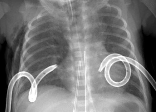 Figure 1:Chest radiograph (Day 1) demonstrates bilateral pigtail catheters
for pneumothorax. Right and left diaphragm at same levels (arrow). An
endotracheal tube is positioned in the trachea, and umbilical arterial and
venous catheters are visualized.
Figure 1:Chest radiograph (Day 1) demonstrates bilateral pigtail catheters
for pneumothorax. Right and left diaphragm at same levels (arrow). An
endotracheal tube is positioned in the trachea, and umbilical arterial and
venous catheters are visualized.
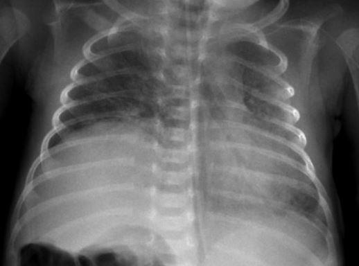 Figure 2:Chest radiograph obtained on day 5 of life demonstrating elevation
of the right hemidiaphragm (Arrow), suggestive of right diaphragmatic
paresis/paralysis. Persistent bilateral hazy pulmonary opacities are present,
consistent with underlying neonatal lung disease. No pneumothorax is
identified.
Figure 2:Chest radiograph obtained on day 5 of life demonstrating elevation
of the right hemidiaphragm (Arrow), suggestive of right diaphragmatic
paresis/paralysis. Persistent bilateral hazy pulmonary opacities are present,
consistent with underlying neonatal lung disease. No pneumothorax is
identified.
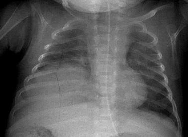 Figure 3:Chest radiograph obtained at 1 month of age demonstrating
persistent elevation of the right hemidiaphragm, consistent with ongoing right
diaphragmatic paresis/paralysis. Low right lung volume is present without
focal consolidation, pleural effusion, or pneumothorax.
Figure 3:Chest radiograph obtained at 1 month of age demonstrating
persistent elevation of the right hemidiaphragm, consistent with ongoing right
diaphragmatic paresis/paralysis. Low right lung volume is present without
focal consolidation, pleural effusion, or pneumothorax.
1. Direct trauma during insertion: The phrenic nerve may be injured during the initial insertion of the chest drain, particularly if the drain is advanced too deeply or directed medially. [3,7]
2. Pressure necrosis: Prolonged compression of the phrenic nerve by a malpositioned chest drain can result in pressure induced ischemia and nerve damage. Ghani et al. hypothesized that chest drain in contact with the phrenic nerve in the pleural apex may cause pressure palsy. [5,9]
3. Hematoma formation: Bleeding at the insertion site or along the drain tract may result in hematoma formation that compresses the phrenic nerve. [9]
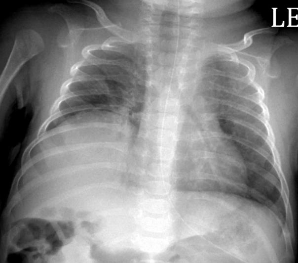 Figure 4:Chest radiograph obtained at 3 months of age demonstrating
persistent elevation of the right hemidiaphragm, consistent with ongoing right
diaphragmatic paresis/paralysis. Low right lung volume persists.
Figure 4:Chest radiograph obtained at 3 months of age demonstrating
persistent elevation of the right hemidiaphragm, consistent with ongoing right
diaphragmatic paresis/paralysis. Low right lung volume persists.
Electrophysiological studies: Phrenic nerve conduction studies and diaphragmatic electromyography may provide additional information regarding the severity of nerve injury and likelihood of recovery. [3,5] These studies can help distinguish between complete nerve transection (poor prognosis) and neurapraxia (better prognosis for recovery).
Conservative management: Mild or asymptomatic cases may be managed conservatively with close monitoring, as spontaneous recovery can occur. [2,3,10] Conservative management includes:
• Continued ventilatory support (invasive or non-invasive) as needed
• Optimization of nutrition to support respiratory muscle function
• Treatment of concurrent pulmonary complications (atelectasis, infection)
• Serial imaging to monitor for recovery
1. Proper insertion technique: Use of the Seldinger technique with pigtail catheters may reduce trauma compared to trocarbased insertion. [1]
2. Optimal positioning: Chest drain should be directed anteriorly and laterally, avoiding medial placement near the spine and superior placement in the apical pleural space. [1,2] [9]
3. Radiographic confirmation: Immediate post-placement radiography should confirm appropriate drain position, with the medial tip at least 1-2 cm from the spine and avoiding the superior mediastinum. [1,2,9]
4. Prompt repositioning: If malposition is identified, the drain should be repositioned or replaced promptly to minimize duration of nerve compression. [5]
5. Clinical vigilance: Clinicians should maintain awareness of this complication and monitor signs of diaphragmatic dysfunction, particularly in infants with difficulty weaning from ventilation after chest drain placement.
Case Report
Diaphragmatic Paralysis Following Pigtail Chest Drain Placement in an Extremely Preterm Infant: A Case Report and Review of Literature
Pan M1, Gaurav Mathur G2, Saju E2, Mubarak A2 and Jain S2*
1John Sealy School of Medicine, The University
of Texas Medical Branch, Galveston, Texas, USA.
2Department of Pediatrics, The University of Texas Medical Branch, Galveston, Texas, USA.
2Department of Pediatrics, The University of Texas Medical Branch, Galveston, Texas, USA.
*Address for Correspondence:Sunil Jain, Department of Pediatrics, The University of
Texas Medical Branch, Galveston, Texas, USA. 301 University Blvd, Galveston, TX, 77550 E-mail Id:
skjain@utmb.edu
Submission: 22 May, 2026
Accepted: 16 September, 2026
Published: 19 September, 2026
Copyright: © 2026 Pan M, et al. This is an open access article
distributed under the Creative Commons Attr-ibution License,
which permits unrestricted use, distribution, and reproduction
in any medium, provided the original work is properly cited.
Keywords:Diaphragmatic Paralysis; Pigtail Chest Drain Insertion;
Pneumothorax; Preterm; Phrenic Nerve Injury.
Abstract
Background: Diaphragmatic paralysis is a rare complication of
chest drain placement in neonates, typically resulting from phrenic
nerve injury related to tube malposition or direct nerve trauma.
Case Presentation: We report a case of right-sided diaphragmatic paralysis following pigtail chest drain insertion for pneumothorax in an extremely preterm male infant born at 26 weeks gestational age. The infant developed pneumothorax requiring chest drain (pig tail) placement with subsequent resolution. Following pig tail removal, imaging confirmed right-sided diaphragmatic paralysis.
Conclusion: This case highlights the importance of proper chest drain (pig tail) positioning and monitoring for this uncommon but significant complication in vulnerable preterm infants. Apicomedial malposition of a pigtail chest drain may result in phrenic nerve injury and diaphragmatic paralysis in extremely preterm neonates. While a definitive causal mechanism cannot be confirmed without direct surgical or electrophysiological evidence, the temporal association, catheter position within the described “danger zone,” and exclusion of alternative etiologies strongly support a causal relationship. Clinicians should ensure chest drain tips are directed laterally and inferiorly, avoiding the apicomedial right hemithorax where the phrenic nerve is most vulnerable. Bedside diaphragmatic ultrasound should be considered following chest drain removal in preterm infants to enable early detection of this complication. A comprehensive review of the literature regarding incidence, mechanisms of injury, diagnostic approaches, and management strategies is provided.
Case Presentation: We report a case of right-sided diaphragmatic paralysis following pigtail chest drain insertion for pneumothorax in an extremely preterm male infant born at 26 weeks gestational age. The infant developed pneumothorax requiring chest drain (pig tail) placement with subsequent resolution. Following pig tail removal, imaging confirmed right-sided diaphragmatic paralysis.
Conclusion: This case highlights the importance of proper chest drain (pig tail) positioning and monitoring for this uncommon but significant complication in vulnerable preterm infants. Apicomedial malposition of a pigtail chest drain may result in phrenic nerve injury and diaphragmatic paralysis in extremely preterm neonates. While a definitive causal mechanism cannot be confirmed without direct surgical or electrophysiological evidence, the temporal association, catheter position within the described “danger zone,” and exclusion of alternative etiologies strongly support a causal relationship. Clinicians should ensure chest drain tips are directed laterally and inferiorly, avoiding the apicomedial right hemithorax where the phrenic nerve is most vulnerable. Bedside diaphragmatic ultrasound should be considered following chest drain removal in preterm infants to enable early detection of this complication. A comprehensive review of the literature regarding incidence, mechanisms of injury, diagnostic approaches, and management strategies is provided.
Introduction
Pneumothorax is a common complication in mechanically
ventilated preterm infants, occurring in 5-7% of very low birth
weight neonates and contributing to increased morbidity and
mortality. [4] Treatment typically involves chest drain placement,
which may be complicated by various adverse events including
infection, bleeding, and organ injury. [2,4] Diaphragmatic paralysis
secondary to phrenic nerve injury from chest drain placement is a
rare but recognized complication in neonates.[5-7] Notably, all
reported cases of chest drain-associated phrenic nerve injury in
neonates have involved traditional large bore intercostal drains
placed by blunt dissection. Pigtail catheters, which are smaller
(typically 8 or 10 French) and inserted percutaneously using the
Seldinger technique, have been increasingly adopted as a less
traumatic alternative for pneumothorax management in preterm
infants. Comparative studies have demonstrated a higher success
rate for pneumothorax resolution with pigtail catheters (96.0% versus
73.7%) and fewer mechanical complications such as subcutaneous
emphysema and drain dislodgement compared with traditional
straight drains. However, pigtail catheters are not without risk —
thoracic organ injuries including lung lobe perforation, pericardial
penetration, and mediastinal injury have been reported, particularly
in extremely preterm infants during resuscitation. [15] Despite this
growing body of evidence on pigtail catheter complications, phrenic
nerve injury resulting in diaphragmatic paralysis has not been
previously reported following pigtail catheter insertion.
The Seldinger technique used for pigtail catheter placement
introduces unique potential mechanisms of phrenic nerve injury
distinct from those of traditional chest drain insertion. These include
direct trauma from the guide wire or dilator during advancement, as
well as sustained compression or traction on the phrenic nerve by the
indwelling catheter if the tip migrates to or is positioned within the
apicomedial pleural space. In extremely preterm neonates, the small
thoracic dimensions and the close anatomic proximity of the phrenic
nerve to the superior vena cava and pericardium may further increase
vulnerability to such injury.
The phrenic nerve originates from cervical nerve roots C3-C5
and courses along the lateral aspect of the mediastinum to innervate
the diaphragm. Its anatomical proximity to the pleural space makes
it vulnerable to injury during chest drain placement, particularly
when tubes are malpositioned too medially or in the superior pleural
space. [1,3,8] Understanding the mechanisms, risk factors, and
management of this complication is essential for clinicians caring for
critically ill neonates.
We report the first case of right-sided diaphragmatic paralysis
following percutaneous pigtail chest drain insertion using the
Seldinger technique in an extremely preterm infant born at 26 weeks’
gestation. This case highlights a previously unreported complication
of pigtail catheter placement and underscores the importance
of careful catheter positioning and post-removal diaphragmatic
assessment in this vulnerable population.
Case Presentation
A male infant was born at 26 weeks’ gestation with a birth weight
of 820 grams (appropriate for gestational age) via emergency cesarean
section for premature prolonged rupture of membranes and footling
breech presentation. Apgar scores were 3, 5, and 8 at 1, 5, and 10
minutes, respectively. The infant required initial resuscitation with
positive pressure ventilation and was admitted to the neonatal
intensive care unit (NICU), where intratracheal surfactant was
administered. Following surfactant administration, the infant had
significant acute deterioration with desaturations and hypotension,
along with decreased breath sounds on the right side. Chest
radiography revealed bilateral pneumothorax. The infant underwent
needle decompression of the pneumothorax followed by Size 6 Fr
pigtail chest drain placement using the Seldinger technique in the
4th intercostal in the anterior to midaxillary line. Follow-up chest
radiography confirmed the position of the tip of catheter lateral and
inferior to apicomedial (danger zone) with appropriate expansion of
the right lung with clinical improvement in saturations and blood
pressure. The chest drain (pig tail) remained in place for 6 days and
was removed after cessation of air leak and continued radiographic
evidence of lung expansion.
In the present case, post-insertion chest radiography did not
demonstrate obvious malposition of the pigtail catheter. Transient
contact between the guidewire or dilator and the phrenic nerve
during the Seldinger insertion technique may cause sufficient
mechanical trauma to produce a neuropraxia, even when the final
catheter position appears acceptable.
Following chest drain removal, chest radiography demonstrated persistent elevation of the right hemidiaphragm. Fluoroscopic examination and diaphragmatic ultrasound revealed paradoxical movement of the right hemidiaphragm during respiration, with the affected hemidiaphragm moving cephalad during inspiration rather than caudally. The diagnosis of right-sided diaphragmatic paralysis secondary to phrenic nerve injury from pigtail chest drain placement was established. The patient was discharged on room air and followed outpatient. At 7 months of age, a chest x-ray was performed, still showing elevated right diaphragm. Patient remained on room air.
Following chest drain removal, chest radiography demonstrated persistent elevation of the right hemidiaphragm. Fluoroscopic examination and diaphragmatic ultrasound revealed paradoxical movement of the right hemidiaphragm during respiration, with the affected hemidiaphragm moving cephalad during inspiration rather than caudally. The diagnosis of right-sided diaphragmatic paralysis secondary to phrenic nerve injury from pigtail chest drain placement was established. The patient was discharged on room air and followed outpatient. At 7 months of age, a chest x-ray was performed, still showing elevated right diaphragm. Patient remained on room air.
We excluded birth traumas leading to unilateral diaphragm as
baby had a footling breech presentation but was born via cesarean
section, which reduces traction forces or the likelihood of a birth related
phrenic nerve injury. Additionally normal pre-drain chest
radiographs with bilateral symmetric diaphragms argue against
birth trauma and congenital diaphragmatic eventration. While
neuromuscular diseases like congenital myopathies, congenital
myasthenic syndromes and spinal muscular atrophy were unlikely
due to normal tone and limb movements, absence of generalized
weakness.[7]
Surgical plication was not pursued in our index case:
Plication is indicated when weaning from ventilation is not
possible or respiratory distress persists beyond the first month of life. If
the infant was successfully weaned, the primary indication was absent
[7] Only 27% of neonates with DP undergo plication, confirming that
conservative management is the predominant approach.[8]
Follow-up after discharge — The case summary should describe:
planned serial imaging (chest radiography and/or diaphragmatic
ultrasound) to monitor for recovery, pulmonary follow-up for BPD
management, and neurodevelopmental follow-up. Diaphragmatic
ultrasound is increasingly preferred over fluoroscopy for serial
monitoring as it avoids radiation exposure and can be performed at
bedside. [7,3]]
Herzog et al. (2025) demonstrated that prematurity is an independent predictor of major complications after plication (41% vs. 19% in term infants), with early gestational age at surgery being an even stronger predictor of morbidity. Complications included prolonged ventilator days and oxygen use at discharge. Spontaneous recovery can occur over months to years, with phrenic nerve regeneration reported up to 3 years after injury. [17]
Gallagher et al. reported a case of successful plication in an ELBW infant, but this was performed only after the infant developed respiratory failure, recurrent atelectasis, and pneumonia but in our index case did not have any of these complications. [18].
Baby did not require surgical intervention as we could wean and extubate the baby on 8 days of life. Spontaneous phrenic nerve recovery with phrenic nerve regeneration takes up to 3 years.
The infant was managed with continued ventilatory support, optimization of nutrition, and close monitoring. Follow-up chest radiography showed persistence of right-sided diaphragmatic paralysis until discharge from the NICU at 3 months of age.
Herzog et al. (2025) demonstrated that prematurity is an independent predictor of major complications after plication (41% vs. 19% in term infants), with early gestational age at surgery being an even stronger predictor of morbidity. Complications included prolonged ventilator days and oxygen use at discharge. Spontaneous recovery can occur over months to years, with phrenic nerve regeneration reported up to 3 years after injury. [17]
Gallagher et al. reported a case of successful plication in an ELBW infant, but this was performed only after the infant developed respiratory failure, recurrent atelectasis, and pneumonia but in our index case did not have any of these complications. [18].
Baby did not require surgical intervention as we could wean and extubate the baby on 8 days of life. Spontaneous phrenic nerve recovery with phrenic nerve regeneration takes up to 3 years.
The infant was managed with continued ventilatory support, optimization of nutrition, and close monitoring. Follow-up chest radiography showed persistence of right-sided diaphragmatic paralysis until discharge from the NICU at 3 months of age.
Review of Literature
Incidence and Epidemiology:
Diaphragmatic paralysis (DP) following chest drain placement
in neonates is a rare complication with limited epidemiological data.
Most evidence comes from case reports and small case series.
[3,5-8] In the context of cardiac surgery, where chest drain is routinely
placed, Ghani et al. reported an incidence of 6.6% (32 of 488 patients)
for right-sided diaphragmatic paralysis in neonates and infants
undergoing cardiac surgery prior to implementation of positioning
guidelines. [9] However, the incidence specifically related to chest
drain placement for pneumothorax in preterm infants appears
to be lower, though precise rates are difficult to establish given
underreporting and diagnostic challenges.
The right hemidiaphragm is more commonly affected than the
left, likely due to anatomical factors and the more frequent placement
of right-sided chest drain. [1,5,6] Preterm infants may be at
particular risk due to their smaller anatomical dimensions, making
precise drain positioning more challenging, and their increased
susceptibility to barotrauma and air leak syndromes requiring chest
drain placement. [2]
Mechanisms of Phrenic Nerve Injury:
The mechanism of phrenic nerve injury from chest drain
placement involves direct mechanical trauma to the nerve. The
phrenic nerve descends along the lateral mediastinum, passing
anterior to the hilum of the lung between the mediastinal pleura and
the pericardium. [3] Several mechanisms have been proposed:1. Direct trauma during insertion: The phrenic nerve may be injured during the initial insertion of the chest drain, particularly if the drain is advanced too deeply or directed medially. [3,7]
2. Pressure necrosis: Prolonged compression of the phrenic nerve by a malpositioned chest drain can result in pressure induced ischemia and nerve damage. Ghani et al. hypothesized that chest drain in contact with the phrenic nerve in the pleural apex may cause pressure palsy. [5,9]
3. Hematoma formation: Bleeding at the insertion site or along the drain tract may result in hematoma formation that compresses the phrenic nerve. [9]
Risk Factors and Tube Positioning:
Multiple studies have identified specific chest drain positions
associated with increased risk of phrenic nerve injury:
Medial positioning: Odita et al. analyzed four cases of neonatal
phrenic nerve paralysis and recommended that the medial end of the
chest drain should be positioned no less than 1 cm from the spine
on frontal chest x-ray. [10] Nahum et al. suggested an even more
conservative distance of at least 2 cm from the vertebrae. [5]
Superior pleural space positioning: Ghani et al. identified a “danger zone” configuration defined as: (1) the chest drains looping apicomedially at the level of the second right intercostal space, and (2) wedging of the chest drain tip against the pericardium. [9] In their cohort, chest drain occupying the right superior pleural space were associated with a 4.22-fold increased risk of DP (95% CI: 1.57-11.33, P <0.05). [9] Following implementation of positioning guidelines to avoid this danger zone, no cases of DP occurred in 43 consecutive patients. [9]
Tube characteristics: The type and size of chest drain may influence risk. Pigtail catheters, while generally considered less traumatic due to their smaller size and softer material, can still cause phrenic nerve injury if malpositioned. [1] Hwang et al. suggested that selection of softer chest drains might ameliorate the problem. [4]
Superior pleural space positioning: Ghani et al. identified a “danger zone” configuration defined as: (1) the chest drains looping apicomedially at the level of the second right intercostal space, and (2) wedging of the chest drain tip against the pericardium. [9] In their cohort, chest drain occupying the right superior pleural space were associated with a 4.22-fold increased risk of DP (95% CI: 1.57-11.33, P <0.05). [9] Following implementation of positioning guidelines to avoid this danger zone, no cases of DP occurred in 43 consecutive patients. [9]
Tube characteristics: The type and size of chest drain may influence risk. Pigtail catheters, while generally considered less traumatic due to their smaller size and softer material, can still cause phrenic nerve injury if malpositioned. [1] Hwang et al. suggested that selection of softer chest drains might ameliorate the problem. [4]
Clinical Presentation and Diagnosis:
DP typically presents with respiratory distress, difficulty weaning
from mechanical ventilation, asymmetric chest wall movement,
pulmonary atelectasis, and recurrent respiratory infections. [3] In
mechanically ventilated preterm infants, the diagnosis may be
suspected when an infant fails to wean from ventilatory support
despite resolution of the primary pulmonary pathology.
Radiographic evaluation:
Chest radiography typically shows elevation of the affected
hemidiaphragm. [3] However, this finding may be subtle in
mechanically ventilated infants with positive pressure support.
Comparison of previous radiographs is essential.
Fluoroscopy:
Fluoroscopic examination has historically been considered the
gold standard for diagnosis, demonstrating paradoxical movement of
the affected hemidiaphragm during respiration (cephalad movement
during inspiration rather than the normal caudal movement).[3]
Ultrasound:
Ultrasound has increasingly replaced fluoroscopy as the preferred
diagnostic modality, as it can be performed at the bedside without
radiation exposure. [3-4] Ultrasound demonstrates absent or
paradoxical diaphragmatic movement and can measure diaphragm
thickness and thickening fraction. Normal diaphragm thickening
during inspiration is typically >20%, while paralyzed diaphragms
show minimal or no thickening. [4]Electrophysiological studies: Phrenic nerve conduction studies and diaphragmatic electromyography may provide additional information regarding the severity of nerve injury and likelihood of recovery. [3,5] These studies can help distinguish between complete nerve transection (poor prognosis) and neurapraxia (better prognosis for recovery).
Management Strategies:
Management of diaphragmatic paralysis depends on clinical
severity and ranges from conservative observation to surgical
intervention. [2-3]Conservative management: Mild or asymptomatic cases may be managed conservatively with close monitoring, as spontaneous recovery can occur. [2,3,10] Conservative management includes:
• Continued ventilatory support (invasive or non-invasive) as needed
• Optimization of nutrition to support respiratory muscle function
• Treatment of concurrent pulmonary complications (atelectasis, infection)
• Serial imaging to monitor for recovery
Surgical diaphragmatic plication: Surgical plication is indicated
for infants with severe respiratory distress, ventilator dependency,
or failure to improve with conservative management. [2,5]
[10] Plication involves suturing the paralyzed hemidiaphragm in a
flattened position to prevent paradoxical movement and improve
respiratory mechanics.
Timing for surgical intervention remains debated. Some centers advocate early plication (within 2-3 weeks) for infants with severe symptoms, while others recommend a trial of conservative management for 4-6 weeks to allow for potential spontaneous recovery. [2,5,10] Rizeq et al. found that diaphragmatic plication performed before 45 days of life was associated with shorter postoperative hospital length of stay compared to later intervention. [8] Conversely, Denamur et al. reported successful conservative management in 63% of neonates and infants with diaphragmatic paralysis following cardiac surgery, with a median ventilation time of 21 days. [14]
Stramrood et al. reported that among 14 newborns with DP due to obstetric phrenic nerve injury, only 4 (29%) recovered spontaneously within 9 days, while 10 (71%) required plication. [13] Those who underwent early plication achieved satisfactory respiratory outcomes in 86% of cases and were successfully extubated within a few days. [13]
Timing for surgical intervention remains debated. Some centers advocate early plication (within 2-3 weeks) for infants with severe symptoms, while others recommend a trial of conservative management for 4-6 weeks to allow for potential spontaneous recovery. [2,5,10] Rizeq et al. found that diaphragmatic plication performed before 45 days of life was associated with shorter postoperative hospital length of stay compared to later intervention. [8] Conversely, Denamur et al. reported successful conservative management in 63% of neonates and infants with diaphragmatic paralysis following cardiac surgery, with a median ventilation time of 21 days. [14]
Stramrood et al. reported that among 14 newborns with DP due to obstetric phrenic nerve injury, only 4 (29%) recovered spontaneously within 9 days, while 10 (71%) required plication. [13] Those who underwent early plication achieved satisfactory respiratory outcomes in 86% of cases and were successfully extubated within a few days. [13]
Prognosis and Recovery:
The prognosis for DP in neonates varies depending on the severity
of nerve injury and underlying patient factors. Spontaneous recovery
occurs in a substantial proportion of cases but may take considerable
time, as phrenic nerve regeneration can require months to years.
[8,10]
Arya et al. reported a case of bilateral diaphragmatic paralysis
in a preterm infant who required 52 days of mechanical ventilation
but eventually made a full recovery. [3] McCool and Tzelepis noted
that in cases of post-traumatic or infectious diaphragmatic paralysis,
spontaneous recovery occurs in approximately two-thirds of patients,
though regeneration of the phrenic nerve may take up to 3 years. [6]
Serial ultrasound monitoring can be used to assess for functional recovery, with improvement in diaphragm thickening fraction correlating with clinical improvement. [12] Summerhill et al. found that among 16 patients with DP, 11 (69%) functionally recovered with a mean recovery time of 14.9 ± 6.1 months. [12]
Factors associated with poor prognosis and need for surgical intervention include:
• Prolonged ventilator dependency (>21 days)
• Severe diaphragm asymmetry (>2 rib segments difference on chest radiography)
• Absence of diaphragm thickening on ultrasound
• Electrophysiological evidence of complete nerve transection
Serial ultrasound monitoring can be used to assess for functional recovery, with improvement in diaphragm thickening fraction correlating with clinical improvement. [12] Summerhill et al. found that among 16 patients with DP, 11 (69%) functionally recovered with a mean recovery time of 14.9 ± 6.1 months. [12]
Factors associated with poor prognosis and need for surgical intervention include:
• Prolonged ventilator dependency (>21 days)
• Severe diaphragm asymmetry (>2 rib segments difference on chest radiography)
• Absence of diaphragm thickening on ultrasound
• Electrophysiological evidence of complete nerve transection
Prevention Strategies:
Prevention of phrenic nerve injury requires meticulous attention
to chest drain placement technique and positioning:1. Proper insertion technique: Use of the Seldinger technique with pigtail catheters may reduce trauma compared to trocarbased insertion. [1]
2. Optimal positioning: Chest drain should be directed anteriorly and laterally, avoiding medial placement near the spine and superior placement in the apical pleural space. [1,2] [9]
3. Radiographic confirmation: Immediate post-placement radiography should confirm appropriate drain position, with the medial tip at least 1-2 cm from the spine and avoiding the superior mediastinum. [1,2,9]
4. Prompt repositioning: If malposition is identified, the drain should be repositioned or replaced promptly to minimize duration of nerve compression. [5]
5. Clinical vigilance: Clinicians should maintain awareness of this complication and monitor signs of diaphragmatic dysfunction, particularly in infants with difficulty weaning from ventilation after chest drain placement.
Discussion
This case illustrates a rare but significant complication of
pig tail chest drain placement in an extremely preterm infant.
The development of DP following pigtail chest drain insertion
for pneumothorax underscores the importance of proper tube
positioning and clinical vigilance in this vulnerable population.
Review of the literature reveals that while DP from chest drain
placement is uncommon, it is well-documented across multiple case
reports and series. The mechanism involves direct phrenic nerve
injury, most commonly from medial or superior malposition of the
chest drain. Recent evidence has identified specific “danger zone”
configurations that significantly increase risk, providing actionable
guidance for prevention. [1]
The diagnostic approach has evolved, with bedside ultrasound increasingly replacing fluoroscopy as the preferred modality for confirming DP and monitoring recovery. [3,4] This is particularly advantageous in the NICU setting, where bedside evaluation minimizes patient transport and radiation exposure.
Management strategies must be individualized based on clinical severity. While spontaneous recovery occurs in a substantial proportion of cases, the timeline for recovery is often prolonged, and some infants require surgical intervention. [2,5,10] The decision regarding timing of diaphragmatic plication remains challenging, balancing the potential for spontaneous recovery against the morbidity of prolonged ventilator dependency. Emerging evidence suggests that earlier surgical intervention (before 45 days) may improve outcomes in infants who ultimately require plication. [2]
Prevention remains the most important strategy. Implementation of positioning guidelines to avoid medial and superior chest drain placement has been shown to eliminate this complication in some series. [1] Radiographic confirmation of appropriate tube position should be routine practice, with prompt repositioning if malposition is identified.
The existing literature on DP from chest drain placement is almost entirely based on traditional (straight) chest tubes, not pigtail catheters specifically. This distinction should be explicitly acknowledged and discussed.
The earliest case reports of neonatal DP from chest drains involved standard intercostal chest tubes in preterm infants. Arya et al. (3) described bilateral DP in a 32-week infant requiring multiple chest drains, with electrophysiological confirmation of phrenic nerve damage and eventual full recovery after 52 days of ventilator dependence. Odita et al. (10) reported four cases of phrenic nerve paralysis from chest tube placement for neonatal pneumothorax, all related to medial malposition of the tube tip, recommending the medial end remain ≥1 cm from the spine on frontal radiograph.
Reed et al. (2016) [16] specifically studied complications of percutaneous pigtail catheters in neonates and infants, identifying lung lobe perforation as the most common injury, along with pericardial and mediastinal injuries — but notably did not report phrenic nerve injury or DP as a recognized complication. This is an important citation to include, as it highlights that DP from pigtail catheters may be underrecognized.
The diagnostic approach has evolved, with bedside ultrasound increasingly replacing fluoroscopy as the preferred modality for confirming DP and monitoring recovery. [3,4] This is particularly advantageous in the NICU setting, where bedside evaluation minimizes patient transport and radiation exposure.
Management strategies must be individualized based on clinical severity. While spontaneous recovery occurs in a substantial proportion of cases, the timeline for recovery is often prolonged, and some infants require surgical intervention. [2,5,10] The decision regarding timing of diaphragmatic plication remains challenging, balancing the potential for spontaneous recovery against the morbidity of prolonged ventilator dependency. Emerging evidence suggests that earlier surgical intervention (before 45 days) may improve outcomes in infants who ultimately require plication. [2]
Prevention remains the most important strategy. Implementation of positioning guidelines to avoid medial and superior chest drain placement has been shown to eliminate this complication in some series. [1] Radiographic confirmation of appropriate tube position should be routine practice, with prompt repositioning if malposition is identified.
The existing literature on DP from chest drain placement is almost entirely based on traditional (straight) chest tubes, not pigtail catheters specifically. This distinction should be explicitly acknowledged and discussed.
The earliest case reports of neonatal DP from chest drains involved standard intercostal chest tubes in preterm infants. Arya et al. (3) described bilateral DP in a 32-week infant requiring multiple chest drains, with electrophysiological confirmation of phrenic nerve damage and eventual full recovery after 52 days of ventilator dependence. Odita et al. (10) reported four cases of phrenic nerve paralysis from chest tube placement for neonatal pneumothorax, all related to medial malposition of the tube tip, recommending the medial end remain ≥1 cm from the spine on frontal radiograph.
Reed et al. (2016) [16] specifically studied complications of percutaneous pigtail catheters in neonates and infants, identifying lung lobe perforation as the most common injury, along with pericardial and mediastinal injuries — but notably did not report phrenic nerve injury or DP as a recognized complication. This is an important citation to include, as it highlights that DP from pigtail catheters may be underrecognized.
Panza et al. (2020) compared pigtail catheters to traditional
chest drains in 44 neonates and found pigtail catheters to be safe and
effective, with fewer complications (no subcutaneous emphysema or
dislodgement), but did not assess for DP as an outcome. [15]
• The present case may therefore represent one of the first reported
instances of DP specifically from a Size 6 Fr pigtail catheter in an
extremely preterm infant, which should be explicitly stated as a novel
contribution. The mechanism of injury — phrenic nerve compression
from medial/superior malposition — is plausibly the same regardless
of drain type, but the smaller caliber and flexibility of pigtail catheters
may theoretically reduce (though clearly not eliminate) this risk.
The largest body of evidence on chest tube–related DP comes
from the postoperative cardiac surgery population, which differs
fundamentally from premature infants with pneumothorax.
• Ghani et al. (2021) — the key study identifying the “danger
zone” configuration — was conducted exclusively in neonates and
infants after cardiac surgery (531 patients), where DP incidence
was 6.6%. In this population, the phrenic nerve may already be
at risk from intraoperative manipulation (retraction, cold injury
from cardioplegia, electrocautery), making it more susceptible to
subsequent pressure injury from a chest tube. This confounding
factor does not exist in premature infants with pneumothorax. [9]
This case contributes to the limited literature on DP following
chest drain placement in extremely preterm infants and reinforces the
need for meticulous technique, radiographic vigilance, and awareness
of this rare but significant complication.
Conclusion
DP is a rare but important complication of chest drain
placement in neonates, resulting from phrenic nerve injury related
to tube malposition or direct trauma. This case report and literature
review highlight the mechanisms of injury, risk factors, diagnostic
approaches, and management strategies for this condition.
Prevention through proper chest drain positioning and radiographic
confirmation is essential. Clinicians should maintain awareness of this
complication and consider DP in the differential diagnosis of infants
with difficulty weaning from mechanical ventilation following chest
drain placement. Management should be individualized based on
clinical severity, with options ranging from conservative observation
to surgical diaphragmatic plication.
Conflict of Interest:
The authors report no conflicts of interest.
Video Clip 1: Right Hemidiaphragm — Paralysis:
Real-time B-mode ultrasound of the right hemidiaphragm
obtained via a subcostal approach using a curvilinear transducer, with
the liver serving as an acoustic window. During quiet respiration and
sniff maneuver, the right hemidiaphragm demonstrates absent caudal
excursion, consistent with diaphragmatic paralysis. No paradoxical
cranial motion is observed. Diaphragmatic thickening fraction is
absent, with no appreciable change in diaphragm thickness between
end-expiration and end-inspiration.
Video Clip 2: Left Hemidiaphragm — Normal Motion:
Real-time B-mode ultrasound of the left hemidiaphragm
obtained via a subcostal approach using a curvilinear transducer, with
the spleen serving as an acoustic window. During quiet respiration
and sniff maneuver, the left hemidiaphragm demonstrates normal
caudal excursion with smooth, symmetric inspiratory descent.
Normal diaphragmatic thickening is observed during inspiration,
consistent with preserved contractile function.Findings are consistent
with isolated right hemidiaphragmatic paralysis with a normally
functioning left hemidiaphragm.
References
Citation
Pan M, Gaurav Mathur G, Saju E, Mubarak A, Jain S. Diaphragmatic Paralysis Following Pigtail Chest Drain Placement in an Extremely Preterm Infant: A Case Report and Review of Literature. J Pediatr Child Care. 2026;12(1): 01.