

Journal of Neurology and Psychology
Download PDF
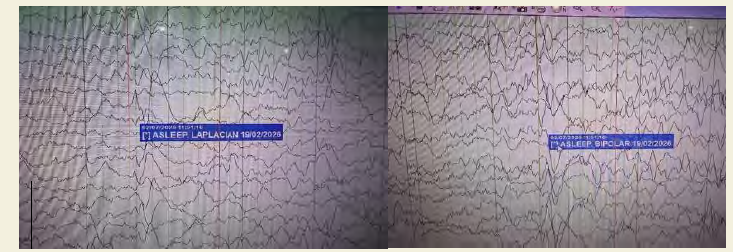 Figure 1A:Evolution of EEG Changes in Anti-NMDAR Encephalitis Over 4 Months.
Background is moderately slow for age, dominated by theta–delta activity. Poorly organized posterior rhythm. Sleep state achieved but architecture is poorly formed. Generalized high-amplitude rhythmic delta activity, intermittent frontal predominance. No definite epileptiform activity.
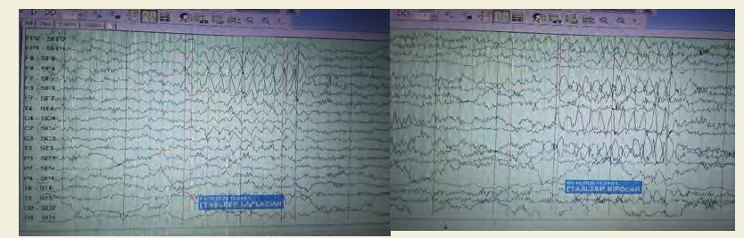 Figure 1B:Background is moderately slow for age, dominated by theta–delta activity. Poorly organized posterior rhythm. Sleep state achieved but architecture is poorly formed. Generalized high-amplitude rhythmic delta activity, intermittent more clear frontal predominance.
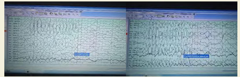 Figure 1C:Background is slow for age, dominated by theta–delta activity. Poorly organized posterior rhythm. Sleep state achieved but architecture is poorly formed. Generalized intermittent high-amplitude non-rhythmic delta activity, in frontal predominance. No definite epileptiform activity.
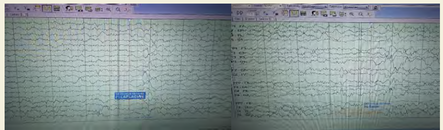 Figure 1D:Background is moderately slow for age, dominated by theta–delta activity. Poorly organized posterior rhythm. Generalized high-amplitude delta activity has decreased as compared with previous EEG record.
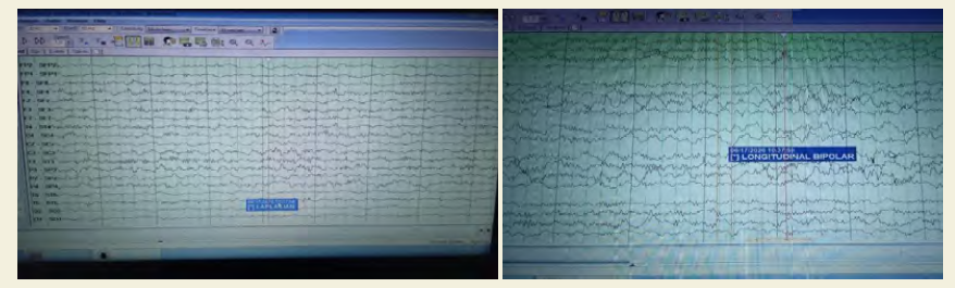 Figure 1E:Background is moderately slow for age, dominated by theta–delta activity. Poorly organized posterior rhythm. Generalized high-amplitude delta activity has almost disappeared.
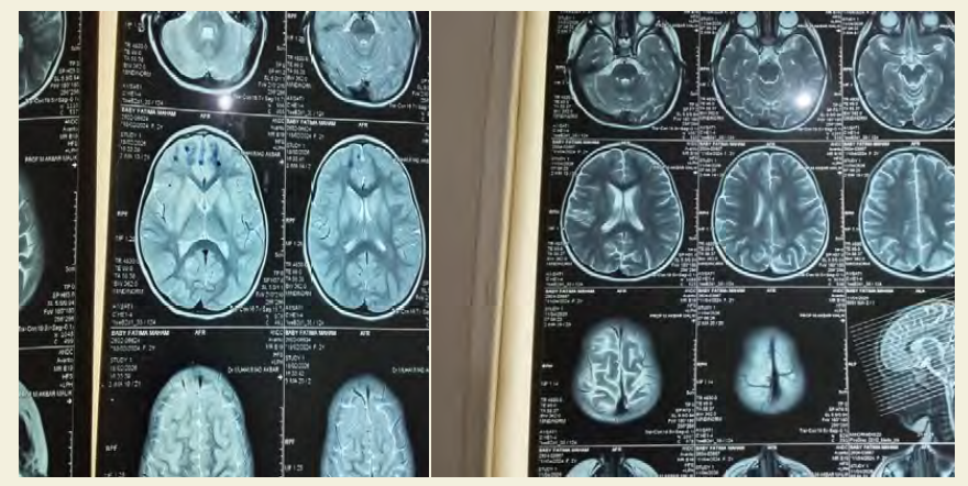 Figure 2A: At admission. Bilateral symmetrical areas of T2/FLAIR hyperintensities in bilateral basal ganglia metabolic/ toxic encephalopathy.
Figure 2B: MRI. After 03 months: Complete radiological resolution of previously noted bilateral basal ganglia signal abnormalities.
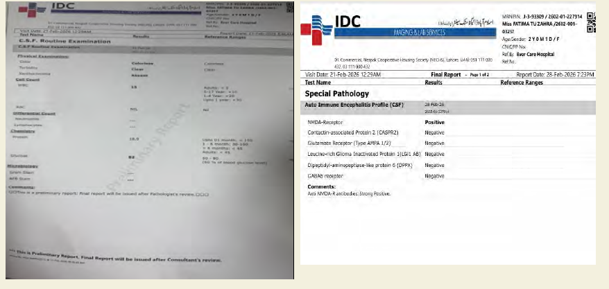 Figure 3:CSF Report on admission.
Case Study
A Twenty Two Months Toddler of Anti-NMDA Receptor Encephalitis Presenting with Total Insomnia for 4 days: The First Toddler Case in World Literature
Muzaffar A, Sadia SJ, Nazar A and Malik MA*
Department of Neuropediatrician the Brain Associates, Multidisciplinary Paediatric Neurosciences Clinic. 218 D. Model Town Lahore-Pakistan
*Address for Correspondence:Muhammad Akbar Malik, Department of Neuropediatrician the Brain Associates. Multidisciplinary Paediatric Neurosciences Clinic. 218 D. Model Town Lahore-Pakistan, E-mail Id: docmalikpk2000@yahoo.co.in
Submission: May 05, 2026
Accepted: July 04, 2026
Published: July 10, 2026
Copyright: ©2026 Muzaffar A, et al. This is an open access article
distributed under the Creative Commons Attribution License, which
permits unrestricted use, distribution, and reproduction in any medium,
provided the original work is properly cited.
Keywords:Autoimmune encephalitis; Cerebrospinal fluid; AntiNMDAR autoimmune encephalitis; Psychosis; Insomnia
Abstract
Anti-N-methyl-D-aspartate receptor encephalitis (anti-NMDARe) is
the most frequent autoimmune encephalitis in children and may cause
long-term sequelae. It is caused by antibodies directed against the RN1
or RN2 subunits of NMDA receptors. It is characterized by a prodromal
phase of unspecific illness with fever that resembles a viral disease.
The prodromal phase is followed by seizures, disturbed consciousness,
psychiatric features, prominent abnormal movements, and autonomic
imbalance. Here, we report a rare case of anti-NMDARe in a 22
months-old girl visited our multidisciplinary neurosciences clinic due to
drug resistant total insomnia and aggression for four days without any
other prodromal symptoms. The cerebrospinal fluid tested positive for
anti-NMDAR antibodies and no association with teratoma or thymoma
was found. The patient was treated with intravenous corticosteroid and
immunoglobulin therapy concomitantly, which resulted in a prompt
clear clinical improvement as she regained the ability to eat, sleep
and social interaction with resumption, without behavioral disturbance
and was discharged after 7 days from the hospital.
Introduction
Anti-NMDA receptor encephalitis (NMDARE) causes
neuropsychiatric symptoms [1,2] resulting in morbidity in 20% [3] and
mortality in 10%. [3,4] It is associated with autoantibodies against the
GluN1 subunit of NMDA receptors on the neuronal surface, leading
to inflammation and dysfunction. This autoimmune encephalitis
develops due to the formation and attachment of IgG, particularly
IgG1 and G3, to NMDA receptor (NR1) subunit with subsequent
internalization of NMDA (glutamate) receptors, reduction of
neuronal Ca influx, and decrease in the receptor-dependent synaptic
currents.[5] Florance et al. [6] reported the first pediatric patient (age
<18 years) with anti-NMDA receptor encephalitis. Although the
clinical manifestation of anti-NMDAR encephalitis in children and
adults was similar, most children did not have underlying tumors. [7]
Anti-NMDAR encephalitis in young children presents with a
distinct, often rapid progression of neuropsychiatric symptoms,
frequently starting with a viral-like prodrome (fever, headache)
followed by behavioral changes (agitation, irritability, tantrums),
seizures, movement disorders (dyskinesia, dystonia), and speech
dysfunction, including mutism. [6,7]
EEG in children with anti-NMDAR encephalitis is abnormal in
80–90% of cases, typically showing non-specific, diffuse, slow-wave
encephalopathy, focal, or multi-focal slowing. While “extreme delta
brush” (EDB) is a signature finding, it is seen in only 6–53% of pediatric
cases. Common findings include background slowing (63–100%)
and epileptiform discharges (33–56%), with 16–52% experiencing
seizures.[10] Brain MRI findings in children with Anti-NMDAR
encephalitis are often normal, reported in 40-60% of pediatric cases
at onset, making it a diagnosis primarily based on clinical symptoms
and CSF antibody testing. When abnormal, findings are typically
non-specific, featuring T2/FLAIR hyperintensities in the cortical/
subcortical areas and hippocampus. [11] CSF analysis in children with
anti-NMDAR encephalitis typically reveals elevated white blood cell
counts (lymphocytic pleocytosis) in >90% of cases, elevated protein
levels, and positive oligoclonal bands (OCBs). The most definitive
diagnostic marker is the detection of anti-NMDAR antibodies in
the CSF, which is more sensitive (100% in some studies) and specific
than serum. [12] The first-line treatment for anti-NMDA receptor
encephalitis is immunotherapy, including methylprednisolone,
intravenous immunoglobulins, and plasma exchange. The secondline
treatment is immunosuppression, typically with rituximab,
azathioprine or cyclophosphamide.[13] Awareness of the disease
and recognition of its symptoms are key to the early diagnosis and
prompt initiation of treatment which may improve outcomes. With
treatment, 75% of patients are estimated to recover completely while
25% may face residual neurodevelopmental impairment or death
[14]. We report a case of a toddler with anti-NMDAR encephalitis
in which the symptom prompting medical evaluation was total
insomnia which has not been previously emphasized in the literature
as a presenting feature of anti-NMDAR encephalitis. On prompt
clinical diagnosis and starting empirical immunosuppressant and
immunomodulating therapy, we were able to discharge her from
hospital on the 7th day of admission.
Case Report
A Twenty-two months girl youngest third child of nonconsanguineous
marriage, with no significant past history and
Background is moderately slow for age, dominated by theta–delta activity. Poorly organized posterior rhythm. Sleep state achieved but architecture is poorly formed. Generalized high-amplitude rhythmic delta activity, intermittent frontal predominance. No definite epileptiform activity.
Figure 2B: MRI. After 03 months: Complete radiological resolution of previously noted bilateral basal ganglia signal abnormalities.
developmentally normal child presented to our multidisciplinary
OPD with total drug-resistant insomnia for last four days. The patient
was a typically developing child born after a full-term uncomplicated
gestation and delivery. She had no prior history of injury, ingestion,
psychosocial stressors, change in appetite, involuntary movements,
or seizures. Three months prior to this presentation, she had been
evaluated by his primary care physician after a three-day febrile
illness and was diagnosed with a viral infection. Since that time, she
had been absolutely well and did not visit any health facility. For the
current illness, she was initially evaluated and treated for insomnia
by various pediatric specialists with risperidone and melatonin, but
her symptoms were worsening over four days and not had slept for a
single hour over last four nights. However, over these four days she
became extremely agitated and started biting herself and her carers.
The family history was negative for neurologic or psychiatric illness.
In the clinic she had episodic outbursts of excessive, inconsolable
crying and distress that were described “as she was having nightmares
while awake”. During these episodes of extreme distress, she was
bursting into tears and requiring physical restraint by family members
against combativeness. The patients’ initial physical examination
revealed normal anthropometry, vital signs, cardiovascular,
pulmonary, and abdominal findings. She did not have dysmorphic
features or a rash. Examination of the cranial nerves, muscle bulk,
tone, reflexes and strength were normal, but she was not straightening
her legs. There was no sign of meningitis on physical examination.
Initial differential for this clinical presentation was broad including
insomnia secondary to pain from corneal abrasion or unidentified
traumatic or infectious source, traumatic brain injury, developmental
disorder with regression, encephalopathy due to acute intoxication,
acute psychosis, postinfectious or parainfectious intravenous and
autoimmune encephalopathy. Spot EEG was done in the clinic and
urgent cerebral MRI was requested. Electroencephalogram (EEG)
was done under sedation with oral ChlorHydrate in the dose of 70mg
per kg, as she was very dysphoric and agitated. Her EEG was nonspecific,
revealing diffuse delta slowing with occasional paroxysmal
high amplitude delta bursts of 1-4 seconds with superadded fast
beta activity but no definite extreme-delta-brush discharges were
documented [Figure 1A]. She was admitted to the general ward under
pediatric neurology services with support from consulting services for
further evaluation and management given his altered mental status
and concern for acute encephalopathy. CSF autoimmune panel was
sent and urgent non-contrast cerebral MRI was requested. Urgent
routine CSF analysis and was normal and after 18 hours of admission
contrast cerebral MRI report was: bilateral symmetrical areas of T2/
FLAIR hyperintensities in bilateral ganglia [Figure 2A]. Possibility of
metabolic/toxic encephalopathy was considered in differentials.
As this toddler was previously absolutely normal, she was empirically initiated intravenous methylprednisolone pulses (30 mg/kg/day) and immunoglobulin (400 mg/kg/day) for five days, considering a provisional diagnosis of autoimmune encephalitis. Eventually, CSF NMDA receptor antibody by fixed cell-based (semiquantitative) assay came back strongly positive [Figure 3]. With prompt immunotherapy with methylprednisolone concomitantly, the child showed marked improvement and after 5 days of pulse therapy she was discharged on oral steroids (hydrocortisone 1mg/ kg 12 hourly) with some intermittent agitation. She was mobile and recognizing all her family members. She is being monitored regularly and after one month of her discharge, her detailed evaluation revealed her normal basal developmental (normal for her chronological age), but her EEG (Figure 1B) was still abnormal. Though she had no apparent convulsions, considering the possibility of subclinical seizures her oral Levetiracetam was continued and being followed monthly.
As this toddler was previously absolutely normal, she was empirically initiated intravenous methylprednisolone pulses (30 mg/kg/day) and immunoglobulin (400 mg/kg/day) for five days, considering a provisional diagnosis of autoimmune encephalitis. Eventually, CSF NMDA receptor antibody by fixed cell-based (semiquantitative) assay came back strongly positive [Figure 3]. With prompt immunotherapy with methylprednisolone concomitantly, the child showed marked improvement and after 5 days of pulse therapy she was discharged on oral steroids (hydrocortisone 1mg/ kg 12 hourly) with some intermittent agitation. She was mobile and recognizing all her family members. She is being monitored regularly and after one month of her discharge, her detailed evaluation revealed her normal basal developmental (normal for her chronological age), but her EEG (Figure 1B) was still abnormal. Though she had no apparent convulsions, considering the possibility of subclinical seizures her oral Levetiracetam was continued and being followed monthly.
On her subsequent monthly visits, we haven’t documented any
residual neurological, behavioral, or sleep-related symptoms after one
month of her discharge from the hospital. However, her asleep EEGs
were abnormal, more or less of the same pattern till 17th JUNE,2026
(5th EEG- (Figure 1E). As, at 4 months of follow-up her EEG was
absolutely normal and clinically and neurologically she was normal
for her age. After this her hydrocortisone is decreased to 0.5 mg/kg/12
hourly, mycophenolate mofetil (MMF) in the dose of 10 mg /kg/12
hourly is commenced and levetiracetam is continued along with
other supportive therapies.
Her long-term management plan is further tapering oral steroids over next 6 months and increasing mycophenolate mofetil to 20 mg/kg/12hourly along with other supportive therapies and close clinical monitoring. Our plan is to continue immunosuppressant/ immunomodulating therapies for two years, if she remains asymptomatic. Due to financial constraints, we will repeat her MRI. Brain and CSF immune panel at one year after the hospital discharges and earlier if she becomes symptomatic
Her long-term management plan is further tapering oral steroids over next 6 months and increasing mycophenolate mofetil to 20 mg/kg/12hourly along with other supportive therapies and close clinical monitoring. Our plan is to continue immunosuppressant/ immunomodulating therapies for two years, if she remains asymptomatic. Due to financial constraints, we will repeat her MRI. Brain and CSF immune panel at one year after the hospital discharges and earlier if she becomes symptomatic
Discussion
The first pediatric case was reported by Höftberger et al. [15]
in 2015, highlighting the clinical features and antibody profiles in
children. The initiating triggers are varied and include viral infections
and tumors [16], which cause the formation of autoantibodies to
glutamate NMDARs, leading to an autoimmune response with
both neurological and neuropsychiatric sequelae. This autoimmune
encephalitis is a disorder with characteristic clinical features that
are predominantly seen in young adults and children with or
without teratomas [6, 17]. Most patients have five stages of clinical
presentation: a prodromal phase, psychotic and/or seizure phase,
unresponsive and/or catatonic phase, hyperkinetic phase, and gradual
recovery phase. [18] In contrast our patient had no other symptom
except total insomnia and agitation. As children with anti-NMDAR
encephalitis have multiple symptoms, and monosymptomatic
cases are present in only 1% of patients that is why anti-NMDAR
encephalitis is unlikely to be a cause of isolated psychosis and is usually
accompanied by seizures [7]. Seizures are a common presentation of
anti-NMDA receptor encephalitis in children and young men [7,19].
Approximately 70% of patients develop seizures, with reported
frequencies of 57% to 82% [20]. Seizures as the sole manifestation
occurs in only 23% of children. Most likely, as in our toddler was
predominantly monosymptomatic of total insomnia without any
complaint of seizures and her diagnosis was being missed.
The diagnosis of anti- NMDAR encephalitis (ANMDARE) is based on clinical suspicion, supported with EEG findings, MRI Brain findings and confirmed by the demonstration of antibodies in the serum or, more sensitively, the cerebrospinal fluid. [7] The electroencephalogram (EEG) is almost always (90–100%) abnormal in cases of ANMDARE and typically shows generalised or pre dominantly frontotemporal slowing [6, 7]. In agreement similar findings of EEG were documented in our toddler.
The diagnosis of anti- NMDAR encephalitis (ANMDARE) is based on clinical suspicion, supported with EEG findings, MRI Brain findings and confirmed by the demonstration of antibodies in the serum or, more sensitively, the cerebrospinal fluid. [7] The electroencephalogram (EEG) is almost always (90–100%) abnormal in cases of ANMDARE and typically shows generalised or pre dominantly frontotemporal slowing [6, 7]. In agreement similar findings of EEG were documented in our toddler.
Epileptogenic abnormalities are less common, seen in 24–50% of
cases, and are possibly more common during the early stage of the
illness [21]. In concordance no definite epileptogenic abnormalities
were present in our patient. The EEG finding of extreme delta brush
(EDB) [17], extreme beta brush [6], and brief rhythmic discharge
[7] have been identified as relatively novel and potential biomarkers
for ANMDARE. However, none of these findings were present in
EEG of our patient. MRI- Brain, an essential tool in diagnosing and
monitoring ANMDARE, can be normal in up to 60% of patients. [22,
23] Common MRI manifestations include cortical T2-hyperintensities
and cortical atrophy, notably in limbic regions. [7,21,24] When MRI
abnormalities are present, they are most found in the temporal,
frontal, and parietal lobes. [25] Though our patient was evaluated
on sixth day of her illness, we documented bilateral basal ganglion
encephalitis. MRI-Brain abnormalities, usually T2-hyperintense
lesions, occur in one-third of children and adults with NMDARE
[1,2]: T2-hippocampal lesions associated with worse outcomes
in adults, but not in children.[26] In agreement, though basal T2-
FLAIR images showed very prominent basal ganglion encephalitis,
our patient was discharged on 7th day of hospitalization and MRIBrain
after three months from discharge showed complete resolution
of basal ganglion hyperintensities. Severe or total insomnia can be the
initial or primary presenting feature of autoimmune encephalitis (AE)
in pediatric patients, even if the classic diagnostic signs are missing
early on, as profound sleep disruption is a known, critical marker of
neuroinflammation in children [6]. This clinical presentation, afebrile
toddler with aggression and EEG findings supported with specific
MRI. Brain findings (basal ganglion encephalitis) in a normally
developing toddler prompted us for the diagnosis of AE and starting
empirical treatment.
Cerebrospinal fluid (CSF) analysis is the cornerstone for
diagnosing pediatric NMDAR encephalitis, with detection of IgG
antibodies against the GluN1 subunit of the NMDAR in the CSF
being the definitive confirmatory test. It is highly sensitive, often
revealing CSF-specific oligoclonal bands (60% of cases), lymphocytic
pleocytosis, and elevated protein, making it more reliable than serum
alone for diagnosis. [13, 17, 27] Cell-based assays (CBA) are the gold
standard for detecting anti-NMDAR antibodies in children with
autoimmune encephalitis, typically testing for IgG against the GluN1
subunit in CSF (highly sensitive), which was positive in our patient
but no other abnormality of CSF was documented. Conversely to
intracellular antigens, cell surface or synaptic proteins antibodies
cannot be detected by standard immunoblot or ELISA and require
the use of cell-based assays. The inclusion of antibodies in the CSF
is vital, since serum testing is less reliable in the diagnosis of anti-
NMDAR AE (14% false negative results) [13,21]
As specific antibody test results can be delayed, treatment
for suspected AE is often given empirically. Early initiation of
immunotherapies has been shown to improve outcomes and reduce
relapses. [1,28] Once the diagnosis is confirmed, the treatment
approach to anti-NMDAR encephalitis generally involves escalation
of immunotherapy alongside teratoma removal where applicable.
[1, 24,29] First line immunotherapies include high-dose steroids,
intravenous immunoglobulins (IVIG) and plasma exchange (PLEX),
sequentially or concomitantly. Second-line immunotherapies, such as
rituximab, azathioprine or cyclophosphamide are used for refractory
cases. Empirically, we used high-dose steroids (Methylprednisolone
30 mg /kg/day single dose and IVIG (400 mg per kg /day for 5 days)
Concomitantly. Our patient responded very well, confirming that
early start and concomitant use of steroids and immunoglobulins are
more effective. This is consistent with a previous report by Titulaer
et al. [4], confirming that these first-line therapies are broad in their
mechanisms of action and can therefore be utilized empirically.
However, to the best our knowledge no toddler in the world literature
with such severe total insomnia has been managed in general ward
and discharged in almost normal health state after seventh day of
admission (11th day after symptoms onset). Therefore, regardless of
antibody status or duration of symptoms, once a diagnosis of possible
or definite AE is made, initiating therapy as soon as possible is
indicated. Children have been reported to recover faster than adults,
usually within six months. [30] Toddlers have a good prognosis, with
full recovery in 67% of patients and no reports of mortality. [31]
In conclusion, because infants and toddlers cannot verbally report hallucinations or delusions, their symptoms are frequently misattributed to viral prodromes, severe temper tantrums, or primary psychiatric issues. Accurate Anti-NMDAR Encephalitis diagnosis in toddlers relies heavily on identifying these non-classical features, as young children rarely present with the textbook adult psychosis. Instead, the presentation is dominated by neurological and developmental red flags. [6] We believe that ANMDARE is probably under reported in infants and toddlers. Early recognition and empirical aggressive immunosuppressive therapies are essential in order to improve outcomes.
In conclusion, because infants and toddlers cannot verbally report hallucinations or delusions, their symptoms are frequently misattributed to viral prodromes, severe temper tantrums, or primary psychiatric issues. Accurate Anti-NMDAR Encephalitis diagnosis in toddlers relies heavily on identifying these non-classical features, as young children rarely present with the textbook adult psychosis. Instead, the presentation is dominated by neurological and developmental red flags. [6] We believe that ANMDARE is probably under reported in infants and toddlers. Early recognition and empirical aggressive immunosuppressive therapies are essential in order to improve outcomes.
Conflict of interest
No potential conflict of interest relevant to this article was reported.
References
Citation
Muzaffar A, Sadia SJ, Nazar A, Malik MA. A Twenty Two Months Toddler of Anti-NMDA Receptor Encephalitis Presenting with Total Insomnia for 4 days: The First Toddler Case in World Literature. J Neurol Psychol. 2026; 12(1): 01.