

Journal of Clinical & Medical Case Reports
Download PDF
In this study, “intraoperative high IOP” was defined as an intraoperative clinical state characterized by shallowing and poor sustainability of the anterior chamber, forward shift of the iris-lens diaphragm, and increased globe tension. This diagnosis was made after excluding other causes such as excessive viscoelastic agent and pupillary block. In some eyes, IOP had been normalized by preoperative medication, laser or anterior-chamber paracentesis, while intraoperative posterior-segment hypertension could still occur. VP was triggered according to these comprehensive intraoperative manifestations instead of a single numerical IOP cutoff.
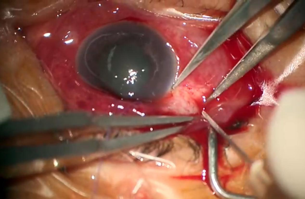 Figure 1:VP in the surgery.
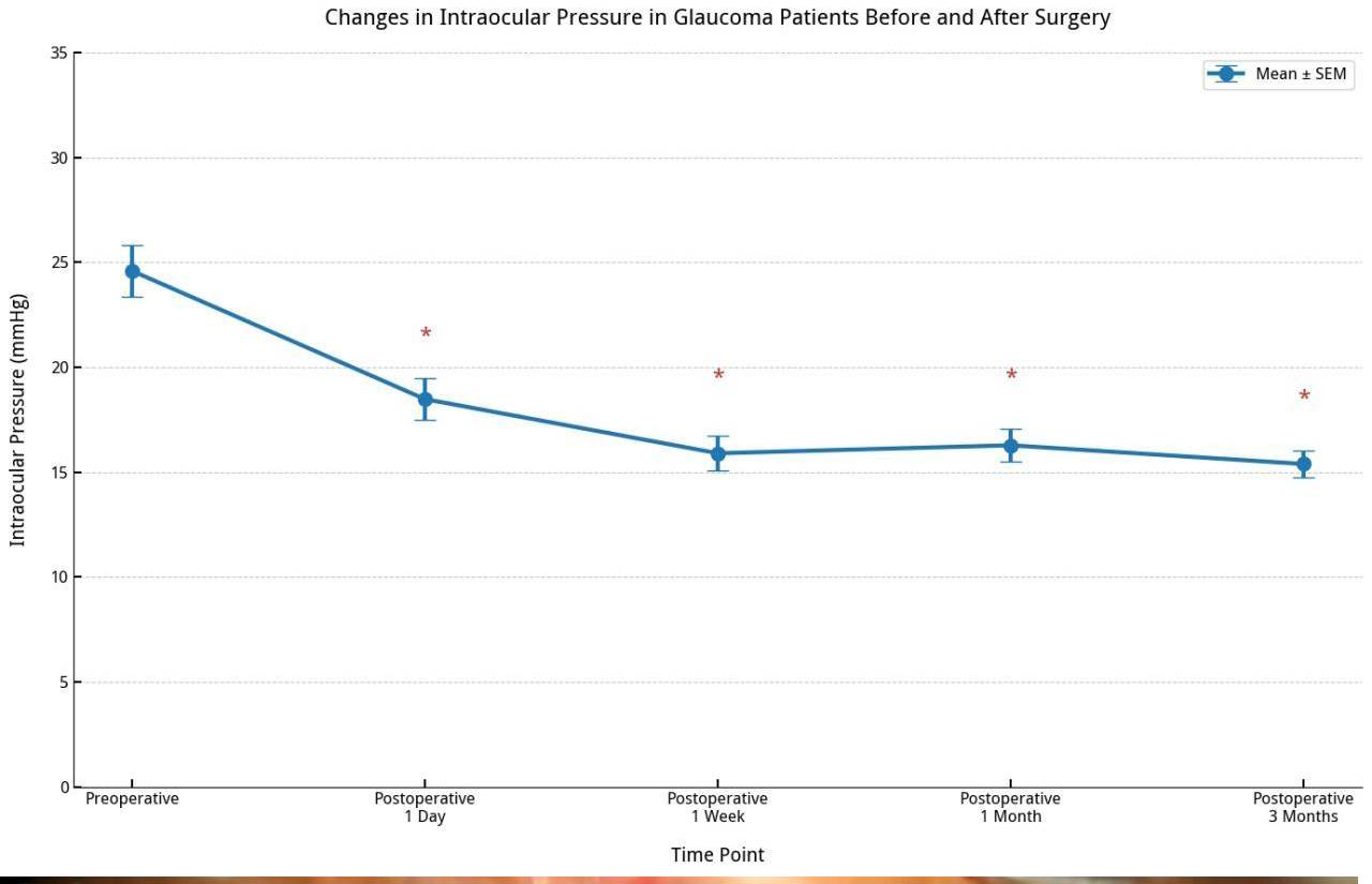 Figure 2:Changes in IOP before and after surgery.
Remarks: * significantly different from preoperative.
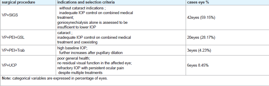 Table 1:Surgical indications and selection criteria for the four glaucoma procedures.
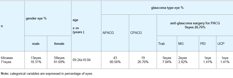 Table 2:General Information of Reserch Subjects.
 Table 3:Ophthalmic Biometric and Intraocular Pressure (Mean±SD).
 Table 4:Preoperative IOP Statistics (n=71eyes).
 Table 5:Success rate of vitreous paracentesis.
 Table 6:Percentage by VP Quadrant n=71eyes).
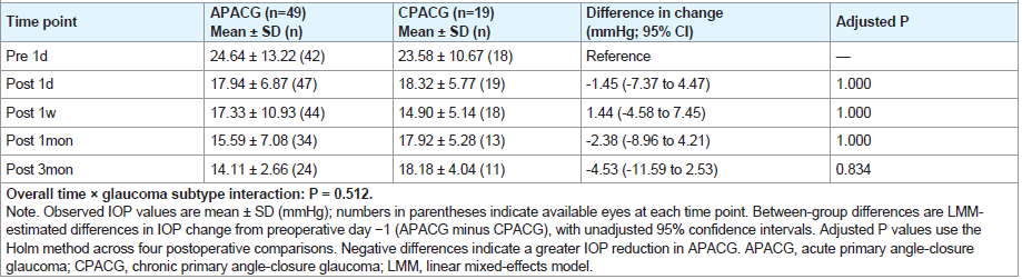 Table 7:Longitudinal IOP and between-group differences by glaucoma subtype.
 Table 8:Longitudinal intraocular pressure changes in the overall cohort and after excluding nine eyes with previous glaucoma surgery.
 Table 9:Longitudinal changes in intraocular pressure after surgery.
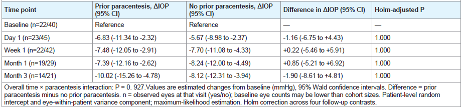 Table 10:Sensitivity analysis of longitudinal IOP changes in all eyes with and without prior anterior chamber paracentesis by linear mixed-effects models.
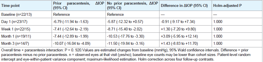 Table 11: Sensitivity analysis of longitudinal IOP changes in APACG eyes with and without prior anterior chamber paracentesis.
A linear mixed-effects model showed no significant timeby- subtype interaction (P = 0.512). Furthermore, no significant between-group differences in IOP reduction were observed at any postoperative time point after Holm correction.
The two eyes with malignant glaucoma were excluded from the inferential subgroup comparison because of the extremely small sample size and were retained for descriptive analysis. Previous glaucoma surgery was considered a clinical characteristic rather than a separate disease subtype, as it may overlap with APACG or CPACG. [Table 7].
Research Article
Clinical Observation of Pars Plana Therapeutic Vitreous Aspiration for Intraoperative High Intraocular Pressure in Primary Angle‑Closure Glaucoma
Ting Ye†¹,², Suju Liu†¹, Xiao Tang¹, Liuzhi Zeng¹*
¹Department of Ophthalmology, Chengdu Integrated TCM & Western
Medicine Hospital, No.18 Wanxiang North Road, High-Tech Zone,
Chengdu, Sichuan Province 610095, China
²Youhua Puhui Eye Hospital, Chengdu, Sichuan Province 610041, China †These authors contributed equally to this work and they are co-first authors
²Youhua Puhui Eye Hospital, Chengdu, Sichuan Province 610041, China †These authors contributed equally to this work and they are co-first authors
Address for Correspondence:Liuzhi Zeng, Department of Ophthalmology, Chengdu Integrated
TCM and Western Medicine Hospital, No.18 Wanxiang North Road, High-Tech Zone, Chengdu, Sichuan Province 610095, China. E-mail Id: 676681961@qq.com
Submission: 09 September, 2026
Accepted: 27 October, 2026
Published: 31 October, 2026
Copyright: © 2026 Zeng L, et al. This is an open access article
distributed under the Creative Commons Attribution License,
which permits unrestricted use, distribution, and reproduction in
any medium, provided the original work is properly cited.
Keywords:Pars plana therapeutic vitreous aspiration; Angle-closure
glaucoma; Intraocular pressure
Abstract
Objective: To investigate the safety and technical feasibility of pars
plana therapeutic vitreous aspiration (VP) combined with glaucoma
surgery for the management of intraoperative high intraocular
pressure (IOP) in primary angle-closure glaucoma (PACG).
Methods: A retrospective study. Patients with PACG who underwent intraoperative VP at the Department of Ophthalmology, Chengdu Integrated TCM& Western Medicine Hospital from July 2022 to January 2026 were enrolled and followed up for 3 months. Preand postoperative IOP, as well as intraoperative and postoperative complications were recorded. The primary endpoint was to evaluate the technical feasibility and improvement of intraoperative surgical conditions of VP during PACG surgery; intraoperative surgical facilitation, postoperative intraocular pressure, complications and visual acuity were designated as secondary outcomes.
Results: A total of 64 patients (71 eyes) were included, consisting of 41 patients (43 eyes, 60.56%) with acute primary angle-closure glaucoma, 16 patients (19 eyes, 26.76%) with chronic primary angleclosure glaucoma, and 7 patients (9 eyes, 12.68%) with PACG after prior glaucoma surgery. The mean IOP preoperative surgery was 24.59 ± 12.34 mmHg. 2. VP: a 1-mL syringe was used to puncture the vitreous cavity at 3.5 mm posterior to the limbus, with a mean aspirated volume of 0.21 ± 0.11 mL. Puncture was performed in the superotemporal quadrant in 35 eyes (49.29%), inferotemporal quadrant in 13 eyes (18.31%), superonasal quadrant in 14 eyes (19.72%), and inferonasal quadrant in 9 eyes (12.68%). The success rate of VP: 65 eyes (91.55%) achieved successful puncture in the first attempt, 5 eyes (7.04%) succeeded on the second attempt, and 1 eye (1.41%) failed all four puncture attempts. 3. Complications: no intraoperative complications such as vitreous hemorrhage or choroidal hemorrhage occurred. Early postoperative complications included hyphema in 2 eyes (2.82%) and vitreous hemorrhage in 1 eye (1.41%); all hemorrhages resolved within one month. Postoperative elevated IOP occurred in 8 eyes (11.27%). The postoperative IOP values at postoperative day 1, week 1, month 1 and month 3 were 18.48 ± 6.98 mmHg, 15.90 ± 8.26 mmHg, 16.28 ± 6.78 mmHg and 15.39 ± 3.64 mmHg, respectively, with model-estimated reductions from baseline and corresponding 95% confidence intervals reported at each follow-up time point.
Conclusion: VP is technically feasible when performed during PACG surgery and may facilitate smooth surgical performance. Limited by the retrospective study design and the absence of quantitative data for intraocular pressure and anterior-chamber depth immediately after the procedure, this study is insufficient to confirm its independent IOP-lowering efficacy. Its clinical value remains to be further validated in comparative studies.
Methods: A retrospective study. Patients with PACG who underwent intraoperative VP at the Department of Ophthalmology, Chengdu Integrated TCM& Western Medicine Hospital from July 2022 to January 2026 were enrolled and followed up for 3 months. Preand postoperative IOP, as well as intraoperative and postoperative complications were recorded. The primary endpoint was to evaluate the technical feasibility and improvement of intraoperative surgical conditions of VP during PACG surgery; intraoperative surgical facilitation, postoperative intraocular pressure, complications and visual acuity were designated as secondary outcomes.
Results: A total of 64 patients (71 eyes) were included, consisting of 41 patients (43 eyes, 60.56%) with acute primary angle-closure glaucoma, 16 patients (19 eyes, 26.76%) with chronic primary angleclosure glaucoma, and 7 patients (9 eyes, 12.68%) with PACG after prior glaucoma surgery. The mean IOP preoperative surgery was 24.59 ± 12.34 mmHg. 2. VP: a 1-mL syringe was used to puncture the vitreous cavity at 3.5 mm posterior to the limbus, with a mean aspirated volume of 0.21 ± 0.11 mL. Puncture was performed in the superotemporal quadrant in 35 eyes (49.29%), inferotemporal quadrant in 13 eyes (18.31%), superonasal quadrant in 14 eyes (19.72%), and inferonasal quadrant in 9 eyes (12.68%). The success rate of VP: 65 eyes (91.55%) achieved successful puncture in the first attempt, 5 eyes (7.04%) succeeded on the second attempt, and 1 eye (1.41%) failed all four puncture attempts. 3. Complications: no intraoperative complications such as vitreous hemorrhage or choroidal hemorrhage occurred. Early postoperative complications included hyphema in 2 eyes (2.82%) and vitreous hemorrhage in 1 eye (1.41%); all hemorrhages resolved within one month. Postoperative elevated IOP occurred in 8 eyes (11.27%). The postoperative IOP values at postoperative day 1, week 1, month 1 and month 3 were 18.48 ± 6.98 mmHg, 15.90 ± 8.26 mmHg, 16.28 ± 6.78 mmHg and 15.39 ± 3.64 mmHg, respectively, with model-estimated reductions from baseline and corresponding 95% confidence intervals reported at each follow-up time point.
Conclusion: VP is technically feasible when performed during PACG surgery and may facilitate smooth surgical performance. Limited by the retrospective study design and the absence of quantitative data for intraocular pressure and anterior-chamber depth immediately after the procedure, this study is insufficient to confirm its independent IOP-lowering efficacy. Its clinical value remains to be further validated in comparative studies.
Introduction
Primary angle-closure glaucoma (PACG) is a group of diseases
characterized by acute or chronic elevation of intraocular pressure
resulting from primary angle closure, leading to optic disc changes
and visual field damage [1]. Acute primary angle-closure glaucoma
(APACG) is a special subtype of PACG classified according to its
acute onset characteristics. Clinically, acute angle-closure glaucoma
predominantly occurs in eyes with markedly bowed iris and narrow
anterior chamber angles, where relative pupillary block is more
pronounced. In the presence of precipitating factors, resistance to
aqueous humor flow from the posterior chamber to the anterior
chamber through the pupil increases, causing elevated posterior
chamber pressure, shallowing of the anterior chamber, and further
elevation of intraocular pressure [2,3]. Due to the unique anatomical
features of PACG-affected eyes and the frequent coexistence of
multiple pathogenic mechanisms [4,5], surgical management
is technically challenging. From July 2022 to January 2026, we
performed pars plana therapeutic vitreous aspiration (VP) prior to
filtering surgery or combined phacoemulsification–trabeculectomy
in PACG eyes at our hospital. This approach yielded satisfactory
outcomes, effectively reducing surgical difficulty and the incidence of
complications. We report our experience as follows.
Subjects and Methods
Subjects:
This was a retrospective study involving 64 patients (71 eyes)
admitted to our hospital with a diagnosis of “angle-closure glaucoma”
from July 2022 to January 2026, who underwent VP during
anti-glaucoma surgery. All surgeries were performed by a single
experienced surgeon.This study was conducted in accordance with
the Declaration of Helsinki and approved by the Ethics Committee
of Chengdu Integrated Traditional Chinese and Western Medicine
Hospital (Approval No. HXKT-065, 2025). Written informed consent
was obtained from the patients and their families.
Methods
Medical and Laser Treatment:
All patients received topical and systemic intraocular pressure lowering
medications upon admission. After medical treatment,
intraocular pressure decreased and corneal clarity was restored, and
laser peripheral iridectomy was performed. In cases where intraocular
pressure remained uncontrolled despite medical therapy and corneal
edema persisted, anterior chamber paracentesis was performed to
reduce corneal edema, followed by laser peripheral iridectomy.In this study, “intraoperative high IOP” was defined as an intraoperative clinical state characterized by shallowing and poor sustainability of the anterior chamber, forward shift of the iris-lens diaphragm, and increased globe tension. This diagnosis was made after excluding other causes such as excessive viscoelastic agent and pupillary block. In some eyes, IOP had been normalized by preoperative medication, laser or anterior-chamber paracentesis, while intraoperative posterior-segment hypertension could still occur. VP was triggered according to these comprehensive intraoperative manifestations instead of a single numerical IOP cutoff.
Surgical Procedures
Based on the surgical principles for PACG outlined in the
Chinese expert consensus on the diagnosis and treatment of primary
glaucoma [1,6], and taking into account the diverse pathogenic
mechanisms in individual patients, the preoperative extent and range
of angle opening, the presence of indications for cataract surgery, and
patient preference, the following surgical approaches were performed
[Table 1]: VP combined with micro-incisional trabeculectomy
and peripheral iridectomy (stab incision glaucoma surgery, SIGS);
VP combined with phacoemulsification with intraocular lens
implantation (PEI) and goniosynechialysis (GSL); VP combined
with ultrasound cycloplasty (UCP); and VP combined with
phacoemulsification, intraocular lens implantation, trabeculectomy,
and peripheral iridectomy (Trab). All patients presented with shallow
anterior chambers and elevated intraocular pressure during surgery,
which increased surgical difficulty and risk. Therefore, after excluding
contraindications such as retinal detachment and suprachoroidal
hemorrhage, VP was performed intraoperatively prior to the primary
procedure.
(1) VP procedure: After topical anesthesia and subconjunctival infiltration anesthesia, a 1- mL syringe fitted with a 26-gauge needle (Sichuan Shuanglu Medical Devices Co., Ltd.) was used. Puncture was performed perpendicularly to the sclera at the pars plana, 3.5 mm posterior to the limbus. Once the needle entered the vitreous cavity to a depth of 2–4 mm, the depth and angle of the needle were adjusted, and gentle intermittent forward-backward movements were applied for trial aspiration. The maximum total intravitreal insertion depth of the needle did not exceed 15 mm. Liquefied vitreous was slowly aspirated, with the volume of vitreous fluid withdrawn ranging from 0.1 to 0.6 mL according to the intraocular pressure. [Figure 1]. Success of VP in this study was defined by intraoperative subjective clinical observations: decreased globe tension, smooth injection of viscoelastic material into the anterior chamber, and visually deepened anterior chamber. No instrumental quantitative measurements of IOP or ACD immediately before and after VP were available. After VP, the anti-glaucoma surgery was commenced. The aspiration sites included the superotemporal, inferotemporal, inferonasal, or superonasal quadrants, which were determined based on operative exposure, prior surgical scars, local anatomical features and the surgeon’s clinical judgment rather than random assignment. If no fluid was obtained on the first attempt, a second puncture was performed at a site 3–4 clock hours away; if the second attempt was unsuccessful, a third puncture was performed at another site 3–4 clock hours away, until vitreous fluid was aspirated with consequent reduction in intraocular pressure and deepening of the anterior chamber. If aspiration remained unsuccessful after the fourth attempt, the procedure was abandoned. After each needle withdrawal, the puncture site was gently compressed with a moist cotton swab against the ocular wall. If intraocular pressure increased and the anterior chamber shallowed during surgery, anterior vitrectomy was performed as necessary.
(2) SIGS in 42 eyes [7] : Following VP, a 2.2 mm corneal puncture knife was inserted through the conjunctiva at a site 1.5 mm posterior to the superior limbus, tunneling into the lamellar sclera and advancing 0.5 mm into the clear cornea before entering the anterior chamber. A scleral punch was then used to excise the inner corneal lip and trabecular tissue, followed by peripheral iridectomy.
(1) VP procedure: After topical anesthesia and subconjunctival infiltration anesthesia, a 1- mL syringe fitted with a 26-gauge needle (Sichuan Shuanglu Medical Devices Co., Ltd.) was used. Puncture was performed perpendicularly to the sclera at the pars plana, 3.5 mm posterior to the limbus. Once the needle entered the vitreous cavity to a depth of 2–4 mm, the depth and angle of the needle were adjusted, and gentle intermittent forward-backward movements were applied for trial aspiration. The maximum total intravitreal insertion depth of the needle did not exceed 15 mm. Liquefied vitreous was slowly aspirated, with the volume of vitreous fluid withdrawn ranging from 0.1 to 0.6 mL according to the intraocular pressure. [Figure 1]. Success of VP in this study was defined by intraoperative subjective clinical observations: decreased globe tension, smooth injection of viscoelastic material into the anterior chamber, and visually deepened anterior chamber. No instrumental quantitative measurements of IOP or ACD immediately before and after VP were available. After VP, the anti-glaucoma surgery was commenced. The aspiration sites included the superotemporal, inferotemporal, inferonasal, or superonasal quadrants, which were determined based on operative exposure, prior surgical scars, local anatomical features and the surgeon’s clinical judgment rather than random assignment. If no fluid was obtained on the first attempt, a second puncture was performed at a site 3–4 clock hours away; if the second attempt was unsuccessful, a third puncture was performed at another site 3–4 clock hours away, until vitreous fluid was aspirated with consequent reduction in intraocular pressure and deepening of the anterior chamber. If aspiration remained unsuccessful after the fourth attempt, the procedure was abandoned. After each needle withdrawal, the puncture site was gently compressed with a moist cotton swab against the ocular wall. If intraocular pressure increased and the anterior chamber shallowed during surgery, anterior vitrectomy was performed as necessary.
(2) SIGS in 42 eyes [7] : Following VP, a 2.2 mm corneal puncture knife was inserted through the conjunctiva at a site 1.5 mm posterior to the superior limbus, tunneling into the lamellar sclera and advancing 0.5 mm into the clear cornea before entering the anterior chamber. A scleral punch was then used to excise the inner corneal lip and trabecular tissue, followed by peripheral iridectomy.
(3) PEI+GSL in 20 eyes: After VP, phacoemulsification and
intraocular lens implantation were performed. Following lens
implantation, viscoelastic was used to support the anterior chamber,
and goniosynechialysis was performed under gonioscopic guidance.
(4) PEI+Trab in 3 eyes: After VP, a 3×4 mm scleral flap was created. Phacoemulsification and intraocular lens implantation were performed, followed by goniosynechialysis under gonioscopic guidance. A trabecular punch was then used beneath the scleral flap to excise a 2×2 mm piece of trabecular tissue, and peripheral iridectomy was performed.
(5) UCP in 6 eyes: After VP, the eye was stabilized using a fixation ring, and the vacuum system was activated. The probe was inserted into the fixation ring, and the procedure was performed under saline bath irrigation while maintaining stable vacuum pressure throughout. Upon treatment initiation, the transducer was activated sequentially in a clockwise manner, with each treatment sector lasting 8 seconds. The treatment encompassed 8 to 10 sectors.
(4) PEI+Trab in 3 eyes: After VP, a 3×4 mm scleral flap was created. Phacoemulsification and intraocular lens implantation were performed, followed by goniosynechialysis under gonioscopic guidance. A trabecular punch was then used beneath the scleral flap to excise a 2×2 mm piece of trabecular tissue, and peripheral iridectomy was performed.
(5) UCP in 6 eyes: After VP, the eye was stabilized using a fixation ring, and the vacuum system was activated. The probe was inserted into the fixation ring, and the procedure was performed under saline bath irrigation while maintaining stable vacuum pressure throughout. Upon treatment initiation, the transducer was activated sequentially in a clockwise manner, with each treatment sector lasting 8 seconds. The treatment encompassed 8 to 10 sectors.
Observation Indicators:
The following data were collected and statistically analyzed:
axial length; preoperative peak intraocular pressure; intraocular
pressure, and anterior chamber depth at 1 day preoperatively, and
at 1 day, 1 week, 1 month, and 3 months postoperatively; as well
as intraoperative and postoperative complications. The primary
endpoint was to evaluate the technical feasibility and improvement of
intraoperative surgical conditions of pars plana therapeutic vitreous
aspiration applied during PACG surgery. Intraoperative surgical
facilitation, postoperative intraocular pressure, complications and
visual acuity were designated as secondary outcomes
Statistical Methods:
Statistical analysis was performed using SPSS version 27.0
software. Longitudinal postoperative intraocular pressure data were
analyzed using linear mixed-effects models incorporating patient level
random intercepts and nested eye-level random intercepts to
account for clustering of bilateral eyes from the same patient. Seven
patients contributed bilateral eyes. All available observations were
included without imputation of missing values. The Holm method
was applied for multiple comparison adjustment. Continuous data
were presented as mean ± standard deviation, along with modelestimated
absolute changes from baseline, 95% confidence intervals
and percentage reductions. Categorical data were reported as counts
and percentages. A P value ≤ 0.05 was considered statistically
significant.
Results
Patient Demographics
A total of 64 patients (71 eyes) were enrolled, including 51 females
(58 eyes, 81.69%) and 13 males (13 eyes, 18.31%). The age at onset
ranged from 48 to 88 years, with a mean of 69.24 ± 10.04 years old.
Among the operated eyes, 43 eyes (60.56%) had acute primary angleclosure
glaucoma (APACG); 19 eyes (26.76%) had chronic primary
angle-closure glaucoma (CPACG); and 9 eyes (12.68%) had undergone
prior anti-glaucoma surgery for angle-closure glaucoma, including
2 eyes (2.82%) with malignant glaucoma (MG) after anti-glaucoma
surgery, 1 eye (1.41%) after phacoemulsification with intraocular lens
implantation (PEI), 5 eyes (7.04%) after trabeculectomy (Trab), and 1
eye (1.41%) after ultrasound cycloplasty (UCP) [Table 2].
Preoperative ocular parameters measured by IOL-Master 700
showed an axial length (AL) ranging from 19.91 mm to 24.42 mm,
with a mean of 23.30 ± 0.88 mm. Ultrasound biomicroscopy (UBM)
revealed a preoperative central anterior chamber depth (ACD)
ranging from 0.78 to 2.87 mm, with a mean of 1.66 ± 0.47 mm. The
preoperative central corneal thickness (CCT) was 583.47 ± 78.61
μm. The presenting intraocular pressure on admission ranged from
18 to 61 mmHg, with a mean of 48.55 ± 10.41 mmHg. A mean of
3.75 types of topical and systemic intraocular pressure-lowering
medications were used. In 48 eyes, corneal clarity was restored after
medical treatment, and laser peripheral iridectomy was performed. In
23 eyes, intraocular pressure remained uncontrolled despite medical
therapy with persistent corneal edema; anterior chamber paracentesis
was performed to reduce corneal edema, followed by laser peripheral
iridectomy. The intraocular pressure on the day before surgery ranged
from 15 to 44 mmHg, with a mean of 24.59 ± 12.34 mmHg [Table 3].
In the study sample, 29 eyes (40.85%) had preoperative intraocular
pressure below 21 mmHg; 15 eyes (21.12%) had pressure between 21
and 30 mmHg; 22 eyes (30.99%) had pressure between 30 and 40
mmHg; and 5 eyes (7.04%) had pressure above 40 mmHg [Table 4].
Following vitreous paracentesis, the surgical procedures
performed in the 71 eyes were as follows: micro-incisional
trabeculectomy in 42 eyes, phacoemulsification combined with
goniosynechialysis in 20 eyes, phacoemulsification combined with
goniosynechialysis and trabeculectomy in 3 eyes, and ultrasound
cycloplasty in 6 eyes [Table 1].
VP Outcomes:
Among the 71 eyes included, successful vitreous aspiration with
deepening of the anterior chamber was achieved in 70 eyes. Of these,
65 eyes (91.55%) were successfully aspirated on the first attempt,
5 eyes (7.04%) on the second attempt, and 1 eye (1.41%) failed to
yield vitreous fluid even after four puncture attempts. The volume of
vitreous fluid aspirated ranged from 0.1 to 0.6 mL, with a mean of
0.21 ± 0.11 mL [Table 5].
Aspiration Sites:
The selection of pars plana entry quadrant was determined based
on operative exposure, prior surgical scars, local anatomical features
and the surgeon’s clinical judgment, rather than random assignment.
The distribution of vitreous aspiration sites across quadrants was as
follows: superotemporal quadrant in 35 eyes (49.30%), inferotemporal
quadrant in 13 eyes (18.31%), superonasal quadrant in 14 eyes
(19.72%), and inferonasal quadrant in 9 eyes (12.68%) [Table 6].
Intraocular Pressure:
We performed a subgroup analysis stratified by glaucoma
subtype, including 49 eyes with acute primary angle-closure glaucoma
(APACG) and 19 eyes with chronic primary angle-closure glaucoma
(CPACG).A linear mixed-effects model showed no significant timeby- subtype interaction (P = 0.512). Furthermore, no significant between-group differences in IOP reduction were observed at any postoperative time point after Holm correction.
The two eyes with malignant glaucoma were excluded from the inferential subgroup comparison because of the extremely small sample size and were retained for descriptive analysis. Previous glaucoma surgery was considered a clinical characteristic rather than a separate disease subtype, as it may overlap with APACG or CPACG. [Table 7].
We performed a sensitivity analysis after excluding the nine eyes
with a history of previous glaucoma surgery. We refitted the same
linear mixed-effects model in the remaining cohort and compared the
estimated postoperative intraocular pressure changes with those from
the full cohort. The sensitivity results are provided in [Table 8]. This
analysis evaluates the robustness of the longitudinal findings to the
inclusion of previously operated eyes. After excluding eyes with prior
glaucoma surgery, the magnitude of IOP reduction at each time point
was broadly similar to that observed in the full cohort, indicating
robustness of the study findings.[Table 9].Subgroup analysis revealed
no significant difference in longitudinal IOP changes between eyes
with and without prior anterior chamber paracentesis (time-by-group
interaction, P = 0.927). Similar results were observed in the sensitivity
analysis restricted to eyes with acute angle-closure glaucoma (P =
0.920).[Table 10,11].
The postoperative intraocular pressures at 1 day, 1 week, 1
month, and 3 months were 18.48 ± 6.98 mmHg, 15.90 ± 8.26 mmHg,
16.28 ± 6.78 mmHg, and 15.39 ± 3.64 mmHg, respectively. Model estimated
reductions from baseline were statistically significant
at all postoperative time points (P < 0.05; [Table 9] [Figure 2].
Postoperative ocular hypertension occurred in 8 eyes (11.27%), which
were treated with intraocular pressure-lowering medications (mean
of 2.3 types). Among these, 3 eyes achieved pressure control, while 5
eyes (7.04%) with uncontrolled intraocular pressure required repeat
anti-glaucoma surgery. Postoperative shallow anterior chamber with
hypotony occurred in 1 eye (1.41%), and shallow anterior chamber
with ocular hypertension occurred in 1 eye (1.41%); both cases were
managed conservatively with satisfactory intraocular pressure control
and restoration of anterior chamber depth.
Complications:
No intraoperative complications such as vitreous hemorrhage
or choroidal hemorrhage occurred in any patient. In the early
postoperative period, hyphema was observed in 2 eyes (2.82%)
and vitreous hemorrhage in 1 eye (1.41%). No clinically significant
change in visual acuity was detected in any of these three eyes during
the follow-up period. Quantitative logMAR visual acuity trajectories
were as follows: one eye with a baseline visual acuity of 1.0 (logMAR
0.0) showed logMAR visual acuities of 0.30, 0.40, 0.22 and 0.10 at
postoperative day 1, week 1, month 1 and month 3, respectively; the
other eye with hyphema had a baseline of no light perception (NLP,
logMAR 3.1) and maintained NLP throughout follow-up; and the
eye with vitreous hemorrhage had a baseline of hand motion (HM,
logMAR 2.6) and retained light perception (LP, logMAR 3.0) from
postoperative day 1 to month 3. All hemorrhages resolved within
1 month postoperatively following symptomatic and supportive
treatment. Postoperative dilated fundus examination was routinely
performed in all eyes to screen for peripheral retinal breaks and
retinal detachment; no retinal tear, retinal detachment, vitreous
incarceration, endophthalmitis, lens injury or macular lesions were
identified in this cohort.
Discussion
Angle-closure glaucoma is characterized by distinctive anatomical
features, including shallow anterior chamber, relatively large and thick
lens, narrow angle entrance, and short axial length, which collectively
contribute to a relatively anterior lens position and crowding of
the anterior segment [2]. In recent years, advances in clinical and
basic research have deepened our understanding of the pathogenic
mechanisms underlying PACG, leading to its classification into the
following five types [1]: (1) Pure pupillary block type: the pupillary
margin is located anterior to the line connecting the scleral spurs, with
increased pupillary block force. When the pupillary block force exceeds
the aqueous pressure in the posterior chamber, aqueous outflow from
the posterior to the anterior chamber through the pupil is obstructed,
resulting in elevated posterior chamber pressure, forward bowing
of the peripheral iris, and consequent narrowing or even closure of
the angle; (2) Plateau iris type [8]: central anterior chamber depth is
normal, but the peripheral iris is thickened with a steep configuration
at the angle entrance, leading to angle narrowing or closure, while
the peripheral iris remains flat without forward bowing; (3) Ciliary
body anterior position type: the ciliary body is positioned anteriorly,
pushing the peripheral iris toward the angle and causing angle
narrowing or closure;(4) Lens-induced type: abnormal positioning
of the lens and its zonules with anterior displacement, reducing
anterior chamber volume and leading to angle closure; (5) Choroidal
expansion type: choroidal vasodilation and increased blood volume
due to various factors (e.g., postural changes, emotional fluctuations,
etc.), resulting in choroidal expansion and thickening, which pushes
the iris–lens diaphragm forward, displacing the iris anteriorly
against the trabecular meshwork, and causing angle narrowing or
closure; (6) Partial ciliary block type: the ciliary ring in PACG eyes
is significantly smaller than that in healthy individuals, and a subset
of patients exhibit partial ciliary block. Consequently, PACG patients
are prone to develop complete ciliary block after trabeculectomy,
and some patients may still experience shallow anterior chamber
and progressive angle closure due to partial ciliary block even after
phacoemulsification combined with goniosynechialysis.
In nearly half of PACG patients in China, multiple pathogenic
mechanisms coexist [4,5,8]. After relieving pupillary block factors,
non-pupillary block factors should be carefully evaluated and
managed accordingly. Clinically, acute angle-closure glaucoma
predominantly occurs in eyes with markedly bowed iris and narrow
angles, where relative pupillary block is more pronounced. In the
presence of precipitating factors, resistance to aqueous humor flow
from the posterior chamber to the anterior chamber through the
pupil increases, leading to elevated posterior chamber pressure.
This predisposes to reverse shallowing of the anterior chamber, and
further elevation of intraocular pressure [2,3].
During an acute angle-closure attack, topical and systemic intraocular pressure-lowering medications should be administered promptly to rapidly reduce intraocular pressure. The choice of surgical procedure should be individualized based on the underlying pathogenic mechanisms, the extent and range of angle opening, the presence of indications for cataract surgery, and the surgical expertise available at the institution. With advances in the understanding of PACG pathogenesis and the development of phacoemulsification techniques, for patients with coexisting cataract who respond poorly to combined intraocular pressure-lowering medications, phacoemulsification with intraocular lens implantation combined with goniosynechialysis under gonioscopic guidance is the preferred approach. In patients with high baseline intraocular pressure that further increases after pupillary dilation, phacoemulsification combined with intraocular lens implantation and trabeculectomy is recommended [9]. For patients without cataract indications who respond poorly to combined medical therapy and are assessed to have insufficient intraocular pressure reduction with goniosynechialysis alone, combined trabeculectomy is advised [10,11]. For PACG patients with poor general health, no residual visual function in the affected eye, and persistent ocular pain with intraocular pressure refractory to multiple treatments, ultrasound cycloplasty (UCP) combined with VP was performed in this study [1,12]. A multicenter, randomized controlled trial involving 155 patients with primary angle closure and 263 patients with PACG across five countries demonstrated that clear lens extraction yielded superior comprehensive outcomes compared with peripheral iridectomy, and it has been recommended as a preferred treatment for PACG [13]. However, given the vast territory of China, the uneven socioeconomic development, and the varying levels of technical expertise and equipment availability among ophthalmic institutions across different regions, the risks associated with lens extraction surgery are higher than those of peripheral iridectomy or laser therapy [14]. Furthermore, patient preferences and needs vary considerably. Therefore, clear lens extraction has not yet been widely adopted. Nevertheless, for PACG cases where non-pupillary block factors—particularly lens-related factors (lensinduced type)—are the predominant pathogenic mechanism, clear lens extraction combined with goniosynechialysis may be considered after comprehensive evaluation based on the institution’s surgical expertise in cataract extraction and patient preference [1,15,16]. Accordingly, in the 71 eyes included in this study, individualized antiglaucoma surgical approaches were adopted after detailed analysis of the underlying pathogenic mechanisms and preoperative and intraoperative findings.
During an acute angle-closure attack, topical and systemic intraocular pressure-lowering medications should be administered promptly to rapidly reduce intraocular pressure. The choice of surgical procedure should be individualized based on the underlying pathogenic mechanisms, the extent and range of angle opening, the presence of indications for cataract surgery, and the surgical expertise available at the institution. With advances in the understanding of PACG pathogenesis and the development of phacoemulsification techniques, for patients with coexisting cataract who respond poorly to combined intraocular pressure-lowering medications, phacoemulsification with intraocular lens implantation combined with goniosynechialysis under gonioscopic guidance is the preferred approach. In patients with high baseline intraocular pressure that further increases after pupillary dilation, phacoemulsification combined with intraocular lens implantation and trabeculectomy is recommended [9]. For patients without cataract indications who respond poorly to combined medical therapy and are assessed to have insufficient intraocular pressure reduction with goniosynechialysis alone, combined trabeculectomy is advised [10,11]. For PACG patients with poor general health, no residual visual function in the affected eye, and persistent ocular pain with intraocular pressure refractory to multiple treatments, ultrasound cycloplasty (UCP) combined with VP was performed in this study [1,12]. A multicenter, randomized controlled trial involving 155 patients with primary angle closure and 263 patients with PACG across five countries demonstrated that clear lens extraction yielded superior comprehensive outcomes compared with peripheral iridectomy, and it has been recommended as a preferred treatment for PACG [13]. However, given the vast territory of China, the uneven socioeconomic development, and the varying levels of technical expertise and equipment availability among ophthalmic institutions across different regions, the risks associated with lens extraction surgery are higher than those of peripheral iridectomy or laser therapy [14]. Furthermore, patient preferences and needs vary considerably. Therefore, clear lens extraction has not yet been widely adopted. Nevertheless, for PACG cases where non-pupillary block factors—particularly lens-related factors (lensinduced type)—are the predominant pathogenic mechanism, clear lens extraction combined with goniosynechialysis may be considered after comprehensive evaluation based on the institution’s surgical expertise in cataract extraction and patient preference [1,15,16]. Accordingly, in the 71 eyes included in this study, individualized antiglaucoma surgical approaches were adopted after detailed analysis of the underlying pathogenic mechanisms and preoperative and intraoperative findings.
In PACG patients, the anatomical characteristics of short
axial length and shallow anterior chamber, together with elevated
intraocular pressure in some cases, increase the difficulty and risk of
filtering surgery or combined phacoemulsification–trabeculectomy,
predisposing to complications such as corneal endothelial injury,
posterior capsular rupture, vitreous prolapse, and explosive
suprachoroidal hemorrhage. Therefore, preoperative control of
intraocular pressure and deepening of the anterior chamber are crucial
for improving surgical success rates and reducing complications.
Preoperative intravenous infusion of mannitol can lower intraocular
pressure through its hyperosmotic dehydrating effect; however, some
patients still present with shallow anterior chamber after infusion.
Moreover, mannitol should be used with caution in elderly patients,
particularly those with hypertension, cardiac insufficiency, renal
insufficiency, or electrolyte disturbances. In addition, excessive
dehydration may cause neuropsychiatric symptoms such as headache
and agitation, leading to poor cooperation during surgery. If
intraocular pressure cannot be controlled or shows no decreasing
trend, anterior chamber paracentesis may be performed prior to
surgery to release aqueous humor and reduce intraocular pressure.
However, in patients with elevated vitreous cavity pressure, lowering
the anterior chamber pressure increases the pressure gradient between
the anterior chamber and the vitreous cavity, resulting in further
shallowing of the anterior chamber [17]. This increases the risk of
radial capsular tears during capsulorrhexis and posterior capsular
rupture [15]; such patients often require combined vitrectomy to
complete the anti-glaucoma procedure, which significantly increases
surgical difficulty, risk, and cost. The use of viscoelastic agents helps
maintain the anterior chamber and protect the corneal endothelium,
but it does not fundamentally deepen the anterior chamber. Overreliance
on viscoelastic agents for anterior chamber maintenance may
also artificially elevate intraocular pressure during surgery, leading to
complications associated with high intraocular pressure.
In 1864, Chandler et al. [18] first proposed vitreous aspiration
during surgery for patients with malignant glaucoma, which could
effectively reduce vitreous pressure. However, as the puncture
was performed beneath a scleral flap using an 18-gauge needle
of relatively large diameter, the procedure carried a considerable
risk of lens injury and excessive vitreous traction, with subsequent
increased risks of postoperative cataract and retinal detachment.
Since then, VP has gradually matured, with improved safety and
expanded indications. In addition to its use in malignant glaucoma,
it has also been applied in anterior segment surgery, as well as for
diagnostic vitreous aspiration in conditions such as endophthalmitis
and intraocular tumors.
This study included 64 PACG patients (71 eyes) with intraoperative posterior-segment hypertension, which needs to be differentiated from pupillary block and classic aqueous misdirection (malignant glaucoma). The core pathogenesis of classic aqueous misdirection/
This study included 64 PACG patients (71 eyes) with intraoperative posterior-segment hypertension, which needs to be differentiated from pupillary block and classic aqueous misdirection (malignant glaucoma). The core pathogenesis of classic aqueous misdirection/
malignant glaucoma is abnormal accumulation of aqueous humor
within the vitreous cavity, leading to vitreous hydration and
expansion that drives sustained forward displacement of the irislens
diaphragm. In contrast, patients in this cohort mainly presented
with acute isolated intraoperative posterior-segment hypertension:
multiple intraoperative factors raised vitreous cavity pressure,
pushing the iris-lens diaphragm forward and resulting in a shallow,
poorly maintained anterior chamber. Some cases were complicated
by concomitant pupillary block but had not developed the complete
pathological cascade of malignant glaucoma; reverse flow of aqueous
humor into the vitreous cavity was not necessarily present. Without
timely intervention, this intraoperative condition carries a potential
risk of further progression to aqueous misdirection.Therefore, in this
study, a 1-mL empty syringe connected with a 26-G needle was used
to perform VP at 3.5 mm posterior to the limbus as an alternative
to conventional vitrectomy. During aspiration, the needle bevel was
oriented upward. After perpendicular insertion into the vitreous cavity
to a depth of 2.0–4.0 mm, gentle small forward-backward movements
were performed for trial aspiration. With slight angulation, further
needle advancement did not exceed 15 mm.We hypothesize several
causes for unsuccessful aspiration of liquefied vitreous during VP:1)
the needle tip fails to reach the liquefied vitreous pocket and remains
within solid non-liquefied vitreous; 2) although the needle enters a
liquefied region, high viscosity of liquefied vitreous or obstruction
by vitreous fibrils impedes fluid withdrawal into the syringe; 3) the
needle tip apposes the vitreous membrane or retinal surface, leading
to valve-like occlusion during aspiration; 4)the needle enters the
canal of Cloquet, yet the volume of liquefied vitreous in this space
is too small to be aspirated. Needle entry into the canal of Cloquet
represents only one hypothesis and cannot be confirmed or refuted
merely based on unsuccessful aspiration. Intraoperative imaging (e.g.,
intraoperative anterior-segment OCT) would be required to verify the
needle tip location.So the needle was then gradually withdrawn with
intermittent aspiration until removal, after which a different puncture
site was selected for a second attempt.The puncture technique used
in this study was similar to that described by Yuan Jiuming et al.
[19]; however, our puncture success rate was 98.59%, compared with
91.2% in the former study. The possible reason for this difference may
be that we used a 26-gauge needle for aspiration, whereas the former
study used a 27-gauge needle. The mean aspirated fluid volume in this
study was only 0.21 ± 0.11 mL. The aspirated fluid was predominantly
liquefied vitreous and may contain a small amount of aqueous humor
that seeped into the pocket; this volume is small relative to the total
vitreous volume. VP targets a localized vitreous fluid pocket rather
than the entire vitreous cavity. The posterior segment of the eye is a
confined space with limited distensibility. Even withdrawal of a small
volume of fluid from this focal pocket can reduce pressure within
this local compartment, alleviate the mechanical forward pushing
force exerted on the iris-lens diaphragm, increase anterior chamber
depth, and facilitate the smooth progression of subsequent surgical
procedures.
Sethi H et al. [20] performed puncture using only a 23-gauge
or 26-gauge needle without a syringe plunger during cataract
surgery in eyes with crowded anterior segment structures, allowing
passive drainage of vitreous fluid and avoiding retinal traction.
However, needle puncture alone requires precise entry into a
vitreous liquefaction cavity for successful drainage; otherwise,
multiple explorations may be necessary. Nossair A.A. et al. [21] used
a 27-gauge needle connected to a 5-mL syringe to aspirate 0.2 mL
of vitreous fluid during phacoemulsification in cataract eyes with
shallow anterior chamber, and reported no complications such as
retinal traction. The authors consider that VP effectively addresses
shallow anterior chamber secondary to short axial length, angle
closure, intumescent cataract, or intraoperative reverse aqueous
flow,thereby reducing corneal endothelial cell injury and lowering the
risk of posterior capsule rupture. Kuriakose T et al. [22] reported that
in eyes with glaucoma with high intraocular pressure or intumescent
cataract, angle-closure glaucoma, or other conditions with potential
aqueous misdirection, vitreous aspiration using a 30-gauge needle
through the pars plana before or during anterior segment surgery
can effectively reduce vitreous pressure. Multiple domestic studies
have also confirmed that in patients with persistent high intraocular
pressure secondary to cataract or glaucoma, performing vitreous
aspiration prior to combined phacoemulsification–trabeculectomy or
trabeculectomy can rapidly reduce intraocular pressure and reform
the anterior chamber. However, the syringes and needles used vary
across studies. For example, Dong Baoying et al.[23] used a 2.5-mL
syringe with a 23-gauge needle for vitreous aspiration, while Huang
Zhijian et al. used a No. 20 blade for entry into the vitreous cavity
followed by aspiration with an 18-gauge needle; Wang Yulei and
Lu Shiheng et al. used a No. 7 needle. In a small number of eyes,
complications such as vitreous hemorrhage occurred [24-27]. In
addition, VP has been used in penetrating keratoplasty (PKP) or
triple procedures combining PKP, extracapsular cataract extraction,
and intraocular lens implantation [28-30] to effectively lower vitreous
pressure. It has also been applied for diagnostic puncture in uveitis,
endophthalmitis [30], and other intraocular infections [31,32], with a low complication rate and favorable safety profile.
The capacity of the syringe and the size of the needle are correlated
with surgical maneuverability; larger sizes are more difficult to
control [33]. Compared with the aforementioned studies, the 1-mL
syringe used in this study generates sufficient negative pressure to
aspirate vitreous fluid, while offering better controllability than the
conventional 2-mL or 5-mL syringes, facilitating the surgeon’s control
over the puncture process and avoiding excessive vitreous traction or
lens injury. In addition, the 26-gauge needle used in this study has an
outer diameter of 0.46 mm and an inner diameter of 0.25 mm, which
allows aspiration of vitreous fluid while minimizing ocular trauma
and vitreous traction, demonstrating a favorable safety profile [34].
In addition, PACG patients are at high risk for malignant glaucoma,
which most commonly occurs after filtering surgery [35,36]. Studies
have suggested that severe postoperative inflammation is one of the
contributing factors [2]. It is proposed that severe postoperative
inflammation may lead to ciliary body congestion and edema, further
narrowing the pre-existing ciliary ring space in PACG patients and
obstructing normal aqueous humor flow. Meanwhile, inflammation
increases exudation, leading to elevated anterior chamber pressure,
which in turn compresses the vitreous and facilitates aqueous entry
into the vitreous cavity, forming a vicious cycle and increasing the
risk of malignant glaucoma [37,38]. Although various treatment
modalities are available, their outcomes are often unsatisfactory,
making prevention particularly important. The most critical
preventive measures include lowering intraocular pressure before
surgery and maintaining anterior chamber stability during surgery
[36]. VP may reduce vitreous cavity volume, lower posterior segment
pressure, and deepen the anterior chamber intraoperatively,
which may theoretically reduce the risk of progression to aqueous
misdirection (malignant glaucoma); however, this potential effect
remains only a hypothesis [39].
Intraoperatively, no complications such as vitreous hemorrhage
or choroidal hemorrhage occurred in any patients. In the early
postoperative period, hyphema was observed in 2 eyes (2.82%) and
vitreous hemorrhage in 1 eye (1.41%). All hemorrhages were absorbed
within 1 month after surgery following symptomatic and supportive
treatment. These intraoperative findings and the low incidence of
severe hemorrhagic complications suggest that VP may reduce
operative difficulty and improve the safety of surgical manipulation
during PACG surgery.
There are several limitations in the present study. First, due to
intraoperative equipment constraints, quantitative measurements of
intraocular pressure and anterior‑chamber depth immediately after
VP could not be obtained.A complete IOP trajectory from admission
→ immediate pre‑VP → immediate post‑VP → postoperative
follow‑up cannot be constructed. We can only compare admission
IOP with the IOP measured one day before surgery to evaluate
the IOP‑lowering effect of preoperative interventions including
medications, anterior‑chamber paracentesis and laser therapy after
admission.In addition, objective tonometry immediately before VP
was unavailable; VP was initiated based on composite intraoperative
clinical signs without a quantitative IOP cutoff value. Only indirect
intraoperative visual signs were available, which cannot directly
verify the physiological changes induced by VP itself.Second, pooled
statistical analysis of postoperative anterior‑chamber depth and
corneal endothelial cell counts was not feasible, as four different
subsequent glaucoma surgical procedures were adopted in this
cohort. For patients receiving VP combined with PEI+GSL or
PEI+Trab, anterior‑chamber depth increased significantly after
lens removal, accompanied by a statistically significant reduction
in corneal endothelial cell density [40]. In eyes treated with VP plus
SIGS or UCP, anterior‑chamber depth only changed mildly under
the influence of filtering efficacy and the effect of UCP ultrasound
energy on the anterior chamber angle[41]; such changes were far less
prominent than those following lens extraction. Given the substantial
inter-procedure differences in impacts on anterior chamber depth
and corneal endothelial cell count, pooled statistical analysis of
preoperative, intraoperative and postoperative anterior chamber
depth and corneal endothelial parameters was not conducted across
all cases. Another important limitation is the absence of a control
group. Because VP was used as an emergency rescue procedure for
intraoperative posterior-segment hypertension in PACG eyes, it
was not clinically or ethically feasible to establish a well-matched
control group undergoing the same glaucoma operations without
VP. Therefore, the present study cannot determine whether VP is
causally associated with intraoperative parameters, operative time,
complications, postoperative IOP, or the need for anterior vitrectomy,
and further comparative studies are required. But, VP alleviates
elevated posterior-segment pressure and stabilizes the anterior
chamber intraoperatively, which may theoretically reduce the risk
of progression to aqueous misdirection (malignant glaucoma).
However, the lack of a control group and limited sample size mean
this remains merely a hypothesis, and the absence of new malignant
glaucoma cases in our follow-up cannot confirm preventive efficacy.
Although intraoperative subjective observations, including
softening of the globe and easy injection of viscoelastic material into
the anterior chamber, may suggest potential intraocular pressure
reduction and anterior-chamber deepening after VP, these are only
indirect intraoperative findings. Without objective quantitative
measurements, they cannot serve as direct evidence to confirm
the independent intraocular-pressure-lowering efficacy of VP and
correlation analysis between aspirated volume and immediate
changes in IOP or ACD cannot be performed. It is expected that
with the popularization of intraoperative anterior-segment OCT and
other imaging techniques in the future, VP can be performed under
image-guided precise localization of liquefied vitreous regions to
further improve surgical safety. Meanwhile, quantitative detection of
intraocular pressure, anterior-chamber depth and vitreous volume
before and after VP can be achieved, providing more robust evidence
for the safety and efficacy of VP in PACG surgery. Furthermore, this
retrospective case series evaluated complications based on available
medical records and fundus documentation. Although dilated fundus
examinations were routinely performed postoperatively for all eyes,
tiny peripheral retinal degeneration or minute retinal breaks might
still be missed. No severe adverse events including retinal tear, retinal
detachment, vitreous incarceration, endophthalmitis, lens injury and
macular lesions were observed in this cohort. Nevertheless, limited
sample size and retrospective data restrict our capacity to rule out
under recording of rare or delayed complications. Furthermore,
quadrant selection was non-random and determined by anatomical
and operative factors, which may introduce potential selection bias.
Meanwhile, we do not claim that VP itself exerts a protective effect
on the corneal endothelium. In the absence of specular microscopy
data of corneal endothelial cell density, this notion remains only a
hypothesis. In addition, our cohort exhibited clinical heterogeneity
between APACG and CPACG cases. Subgroup sample sizes were
limited, which may reduce statistical power to detect subtle intergroup
differences in IOP trajectories. The two malignant glaucoma eyes
were limited to descriptive analysis without inferential comparison.
Prior glaucoma surgery was treated as a clinical characteristic rather
than a separate diagnostic subgroup and may introduce confounding
effects. Given the heterogeneity of surgical procedures performed in
the included eyes, postoperative visual acuity varied substantially.
Therefore, we only reported the visual acuity outcomes of the three
eyes that developed hyphema or vitreous hemorrhage. Despite the
occurrence of hyphema or vitreous hemorrhage in these three eyes,
no clinically significant change in visual acuity was observed during
the follow-up period. Accordingly, the overall safety profile of VP
cannot be fully established in the present study, and its long-term
complication risks require further validation in larger prospective
studies with extended follow-up.
In conclusion, pars plana therapeutic vitreous aspiration
performed with a 1-mL syringe and a 26-G needle is technically
feasible and can facilitate surgical procedures during PACG glaucoma
surgery. Nevertheless, constrained by the inherent limitations of this
retrospective uncontrolled study, the absence of objective immediate
post-VP measurements, and heterogeneity of subsequent glaucoma
surgeries, the independent intraocular-pressure-lowering effect of VP
cannot be confirmed in our study. Based on observations from this
case series, this technique demonstrates certain clinical application
potential. Its definite clinical value awaits further evaluation in
subsequent prospective comparative studies.
References
Citation
Ting Ye, S Liu, X Tang, L Zeng. Clinical Observation of Pars Plana Therapeutic Vitreous Aspiration for Intraoperative High Intraocular Pressure in Primary Angle‑Closure Glaucoma. J Clin Med Case Reports. 2026;10(1): 12.