

Journal of Clinical and Investigative Dermatology
Download PDF
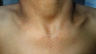 Figure 1A:Lacy reticulate hyperpigmentation on the upper chest and anterior
aspect of the neck.
Figure 1A:Lacy reticulate hyperpigmentation on the upper chest and anterior
aspect of the neck.
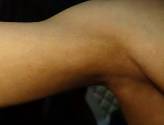 Figure 1B:Larger areas of reticulate, partially confluent hyperpigmentation
admixed with hypopigmented macules over the right axilla and upper arm.
Figure 1B:Larger areas of reticulate, partially confluent hyperpigmentation
admixed with hypopigmented macules over the right axilla and upper arm.
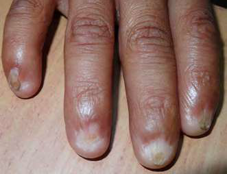 Figure 1C:Near-complete loss of nails of the third and fourth digits of the
right hand (anonychia) and partial loss of nails of the second and fifth digits
(micronychia), with dorsal pterygia and wrinkling of the skin over the distal
phalanges.
Figure 1C:Near-complete loss of nails of the third and fourth digits of the
right hand (anonychia) and partial loss of nails of the second and fifth digits
(micronychia), with dorsal pterygia and wrinkling of the skin over the distal
phalanges.
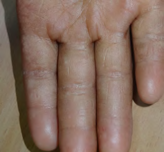 Figure 1D:Right palmar hyperkeratosis with superimposed guttate
hypomelanotic macules and loss of dermatoglyphics (adermatoglyphia).
Figure 1D:Right palmar hyperkeratosis with superimposed guttate
hypomelanotic macules and loss of dermatoglyphics (adermatoglyphia).
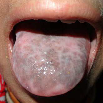 Figure 2A:Confetti-like hypopigmented macules with lacy reticulated
hyperpigmentation over the submental area and anterior aspect of the neck.
Figure 2A:Confetti-like hypopigmented macules with lacy reticulated
hyperpigmentation over the submental area and anterior aspect of the neck.
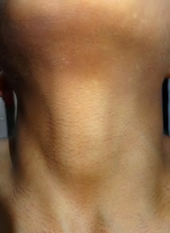 Figure 2B:Confetti-like hypopigmented macules with lacy reticulated
hyperpigmentation over the submental area and anterior aspect of the neck.
Figure 2B:Confetti-like hypopigmented macules with lacy reticulated
hyperpigmentation over the submental area and anterior aspect of the neck.
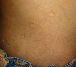 Figure 2C:Three atrophic scars in an oblique linear distribution over the
right flank.
Figure 2C:Three atrophic scars in an oblique linear distribution over the
right flank.
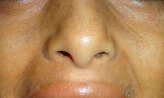 Figure 2D:Fine, lacy, reticulated pigmentation over the nose, cheeks, and
upper lip.
Figure 2D:Fine, lacy, reticulated pigmentation over the nose, cheeks, and
upper lip.
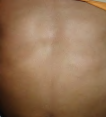 Figure 3A:Lacy reticulate pigmentation over the chest and abdomen, along
with two atrophic macules on the right lower abdomen.
Figure 3A:Lacy reticulate pigmentation over the chest and abdomen, along
with two atrophic macules on the right lower abdomen.
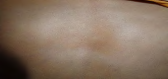 Figure 3B:Lacy reticulate pigmentation over the back, producing the
impression of a reticulated atrophic lesion.
Figure 3B:Lacy reticulate pigmentation over the back, producing the
impression of a reticulated atrophic lesion.
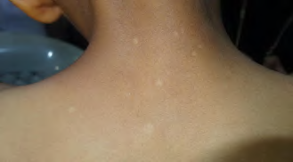 Figure 3C:Large hypopigmented macules over the upper back and nape
of the neck.
Figure 3C:Large hypopigmented macules over the upper back and nape
of the neck.
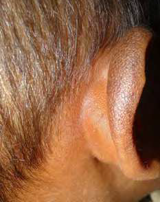 Figure 3D:Dense reticulate hyperpigmentation of the right helix accompanied
by premature graying of the scalp hair.
Figure 3D:Dense reticulate hyperpigmentation of the right helix accompanied
by premature graying of the scalp hair.
Case Report
Atypical Cutaneous Manifestations of Dyskeratosis Congenita: A Case Report from Yemen
Alshami MA1*, Alshami AM2, Alshami HM1 and Lutf RM1
1Department of Dermatology, Faculty of Medicine and Medical Sciences,
Sana’a University, Sana’a, Yemen
2Department of Conservative Dentistry, Faculty of Dentistry, Sana’a University, Sana’a, Yemen
2Department of Conservative Dentistry, Faculty of Dentistry, Sana’a University, Sana’a, Yemen
*Address for Correspondence:Mohammad Ali Alshami, Department of Dermatology, Faculty of
Medicine and Medical Sciences, Sana’a University, Sana’a 1064, Yemen. E-mail Id: mohammadalshami62@gmail.com
Submission: 16 August, 2026
Accepted: 29 September, 2026
Published: 06 October, 2026
Copyright: © 2026 Alshami MA, et al. This is an open access
article distributed under the Creative Commons Attribution License,
which permits unrestricted use, distribution, and reproduction in any
medium, provided the original work is properly cited.
Abstract
Dyskeratosis congenita is a rare genodermatosis characterized
by a classic triad of reticulate skin pigmentation, nail dystrophy, and
mucosal leukoplakia, along with a predisposition to malignancy. We
report the case of a 15-year-old boy who presented to our dermatology
clinic with the characteristic triad and several rare findings.
Unfortunately, genetic testing was not available, so the diagnosis was
based solely on clinical findings. To the best of our knowledge, only one
case with the additional findings of atrophic scars, reticulated atrophy,
and hypopigmented macules has been reported in the literature to
date, and no cases of reticulate pigmentation of the helix have yet
been described.
Introduction
Dyskeratosis congenita (DC), also known as Zinsser–Engman–
Cole syndrome, is a rare multisystem genodermatosis with an
estimated incidence of approximately 1 per million people, it was
originally described by Zinsser in 1906.[1,2] DC is considered the
archetypal telomere biology disorder (TBD) and is characterized by
the classic triad of oral leukoplakia, nail dystrophy, and reticulate skin
pigmentation.[3] Although X-linked DC due to DKC1 mutations is
an important form, DC/TBDs have multiple inheritance patterns,
including autosomal dominant and autosomal recessive forms.
Most cases are associated with defects in the DKC1 gene, which
encodes the dyskerin protein involved in telomere maintenance.[4]
DC is most commonly inherited in an X-linked recessive disorder;
therefore, males are affected approximately three times more often
than females. Approximately 80% of DC cases exhibit associated bone
marrow failure.[5]
Case report
A 15-year-old boy presented to the dermatology clinic with
dystrophic nails and dyspigmentation of the skin and tongue. He was
short for his age, and was wearing a diaper due to a urethral stricture
that had recently been surgically corrected.
On cutaneous examination, a lacy, reticulated hyperpigmentation pattern was identified on the neck, upper chest, and upper abdomen, occasionally admixed with confetti-like hypopigmented macules. In addition, all 20 nails were dystrophic, hypoplastic, or absent, with dorsal pterygia. The patient’s tongue was covered with white reticulated plaques [Figure 1A-D]. Ophthalmologic examination revealed epiphora (excessive tearing) of the left eye.
On cutaneous examination, a lacy, reticulated hyperpigmentation pattern was identified on the neck, upper chest, and upper abdomen, occasionally admixed with confetti-like hypopigmented macules. In addition, all 20 nails were dystrophic, hypoplastic, or absent, with dorsal pterygia. The patient’s tongue was covered with white reticulated plaques [Figure 1A-D]. Ophthalmologic examination revealed epiphora (excessive tearing) of the left eye.
The patient also exhibited multiple oval-shaped atrophic scars in
an oblique linear distribution over the right flank [Figure 2C]. Larger
hypopigmented macules were present on the upper back and nape of
the neck, with smaller ones on the anterior neck [Figure 2B], [Figure 3C]. The patient also had adermatoglyphia of both palms and early
graying of the scalp hair [Figure 1D &3D].
The initial differential diagnoses included reticulate
The initial differential diagnoses included reticulate
acropigmentation of Kitamura, dyschromatosis symmetrica
hereditaria, Naegeli–Franceschetti–Jadassohn syndrome,
dermatopathia pigmentosa reticularis, and Dowling–Degos disease.
Although mucosal lichen planus was considered a differential
diagnosis of the oral lesions, the other cutaneous findings were
inconsistent with this diagnosis. Based on the characteristic clinical
findings, the patient was diagnosed with DC. The patient fulfilled the
diagnostic criteria for DC, meeting three major criteria—reticulate
pigmentation, nail dystrophy, and oral leukoplakia—and three minor
criteria—early graying of hair, urethral stricture, and short stature
[6,7].
Laboratory investigations revealed a white blood cell count of 20.1 × 10⁹/L, with 75.8% neutrophils and 17.9% lymphocytes, a platelet count of 485 × 109/L, and a hemoglobin level of 10.9 g/dL. A punch biopsy was declined by the patient’s parent. These routine laboratory investigations did not indicate bone marrow involvement,
Laboratory investigations revealed a white blood cell count of 20.1 × 10⁹/L, with 75.8% neutrophils and 17.9% lymphocytes, a platelet count of 485 × 109/L, and a hemoglobin level of 10.9 g/dL. A punch biopsy was declined by the patient’s parent. These routine laboratory investigations did not indicate bone marrow involvement,
which occurs in up to 80% of cases and may progress to bone
marrow failure, necessitating hematopoietic cell transplantation. This
apparent leukocytosis with neutrophilia was considered to be due to
bacterial infection, while the anemia was attributed to malnutrition,
which is common in our country. Hematological consultation was
advised, but the parents refused a bone marrow aspiration/biopsy.
No abnormalities were identified in the neurological, pulmonary, orthopedic, or gastrointestinal systems. A genetic test for a mutation in the CTC1, DKC1, TERC, TERT, TINF2, NHP2, NOP10 and WRAP53 genes, all essential for proper telomere function, would have confirmed the diagnosis of DC; however, this test was unavailable at our institution. The patient was scheduled for follow-up visits every 6 months but was lost to follow up.
No abnormalities were identified in the neurological, pulmonary, orthopedic, or gastrointestinal systems. A genetic test for a mutation in the CTC1, DKC1, TERC, TERT, TINF2, NHP2, NOP10 and WRAP53 genes, all essential for proper telomere function, would have confirmed the diagnosis of DC; however, this test was unavailable at our institution. The patient was scheduled for follow-up visits every 6 months but was lost to follow up.
Discussion
The present case expands the clinical spectrum of DC by
demonstrating several uncommon cutaneous manifestations in
addition to the characteristic mucocutaneous triad.[8] Specifically,
the patient exhibited multiple oval-shaped atrophic scars arranged
in an oblique linear distribution over the right flank, a finding that
has been described only once previously, by Milgrom in 1964. Larger
hypopigmented macules were also present on the upper back and
nape, with smaller macules on the anterior neck and submental region;
these findings have been reported only once previously, by Ward et al.
in 2017.[9]This case is unique in that the patient exhibited not only
the typical mucocutaneous triad but also several rare manifestations.
To the best of our knowledge, atrophic scars, reticulated atrophy
[Figure 3B], and hypopigmented macules have each been described
only once previously in the literature. Furthermore, reticulate
pigmentation of the helix, as observed in this patient, has, to the best
of our knowledge, not been previously reported, as a Medline search
revealed no results.
References
Citation
Alshami MA, Alshami AM, Alshami HM, Lutf RM. Atypical Cutaneous Manifestations of Dyskeratosis Congenita: A Case Report from Yemen. J Clin Investigat Dermatol. 2026;14(1): 1