

Journal of Clinical and Investigative Dermatology
Download PDF
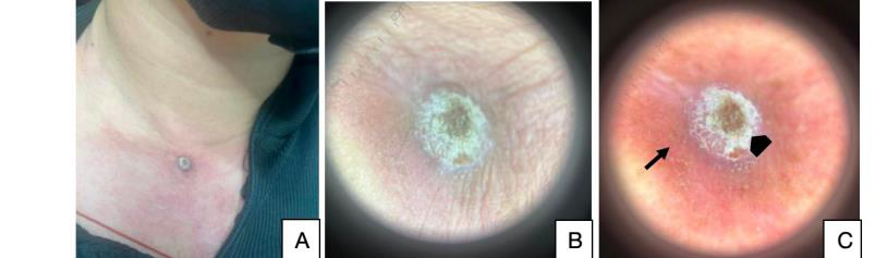 Figure 1:Clinical and dermoscopic features of the lesion. (A) Solitary 2 × 2–cm erythematous crateriform nodule with a central keratin-filled crater arising on an erythematous plaque over the upper anterior chest prior to excision. (B) Nonpolarized dermoscopy. (C) Polarized dermoscopy showing white-to-red structureless areas (black arrow) and irregular polymorphous vessels (arrowhead).
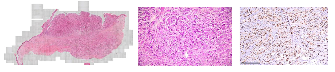 Figure 2:Histopathologic and immunohistochemical findings. (A) Low-power view (hematoxylin-eosin [H&E], ×100) demonstrating epidermal ulceration overlying infiltrative islands of atypical, acantholytic-appearing epithelial cells within the dermis. (B) High-power view (H&E, ×400) demonstrating marked nuclear pleomorphism, hyperchromasia, and frequent mitotic figures. (C) Immunohistochemical staining demonstrating diffuse nuclear positivity for GATA-binding protein 3 (GATA3), supporting metastatic breast carcinoma.
Case Report
Keratoacanthoma-like Cutaneous Metastasis from Invasive Ductal Carcinoma of the Breast: A Case Report
Princess MFP *, Mendoza A and Villafuerte L
Department of Dermatology, Jose R. Reyes Memorial Medical Center, Manila, Philippines
*Address for Correspondence:Princess Marie F. Paredes, Department of Dermatology Jose R.
Reyes Memorial Medical Center San Lazaro Compound, Rizal Avenue, Sta. Cruz Manila 1003, Philippines. E-mail Id: paredescess@gmail.com
Submission: 07 July, 2026
Accepted: 04 August, 2026
Published: 08 August, 2026
Copyright: © 2026 Princess MFP, et al. This is an open access
article distributed under the Creative Commons Attribution License,
which permits unrestricted use, distribution, and reproduction in any
medium, provided the original work is properly cited.
Abbreviation and Acronym List:
CK7 – Cytokeratin 7; CK20 – Cytokeratin 20; ER – Estrogen
receptor; GATA3 – GATA-binding protein 3; HER2 – Human
epidermal growth factor receptor 2; KA – Keratoacanthoma; KA-like
– Keratoacanthoma-like; PR – Progesterone receptor.
Introduction
Cutaneous metastases occur in approximately 0.5%–10.4% of
patients with internal malignancies, and generally signify advanced
disease with an unfavorable prognosis.[1] Breast carcinoma is the
most common internal malignancy associated with cutaneous
metastases in women, whereas lung carcinoma is the most common
primary source in men.[2] Cutaneous metastases from breast
carcinoma exhibit diverse clinical morphologies, including papules,
nodules, erysipeloid, telangiectatic, ulcerative, zosteriform, and
sclerodermoid lesions.[3] Among the rarest clinical presentations is
keratoacanthoma-like (KA-like) cutaneous metastasis, which may
closely mimic primary cutaneous squamous neoplasms and delay
recognition of metastatic disease.
KA-like cutaneous metastases are exceedingly rare and have
been reported in association with lung, esophageal, laryngeal, renal,
prostatic, and breast carcinomas, with lung carcinoma representing
the most frequently reported primary source. [4,5] Because these
lesions closely resemble primary cutaneous tumors on clinical
examination, accurate diagnosis requires careful clinicopathologic
correlation supported by immunohistochemical evaluation.
Herein, we report a rare case of KA-like cutaneous metastasis
arising from invasive ductal carcinoma of the breast in a 40-yearold
Filipino woman. This case highlights the diagnostic challenge
posed by this uncommon morphologic variant and underscores the
importance of integrating clinical history with histopathologic and
immunohistochemical findings to establish the correct diagnosis and
guide appropriate oncologic management.
Case Report:
A 40-year-old Filipino woman presented with a rapidly
enlarging crateriform lesion on the anterior chest, arising against a
background of two metachronous, contralateral primary invasive
ductal carcinomas of the breast diagnosed three years apart. Three
years earlier, she had undergone modified radical mastectomy with
axillary lymph node dissection and breast reconstruction for stage
IIIC right-sided invasive ductal carcinoma. Immunohistochemistry
demonstrated estrogen receptor (ER)-negative, progesterone
receptor (PR)-negative, and human epidermal growth factor receptor
2 (HER2)-negative disease. She subsequently received adjuvant
chemotherapy with doxorubicin and cyclophosphamide, followed
by 33 sessions of radiotherapy to the right breast.
During follow-up, a new mass in the contralateral (left) breast
was identified and confirmed by fine-needle aspiration biopsy. This
second tumor was ER-negative, PR-negative, and HER2-positive — a
receptor profile discordant from the first, HER2-negative carcinoma
— and was therefore classified as a second, biologically independent
primary rather than a local recurrence or contralateral metastasis of
the original right-sided tumor, in which the receptor profile would
be expected to be preserved. In view of the HER2-positive status,
trastuzumab was subsequently added to her treatment regimen.
Two months before consultation, she noted a solitary
erythematous papule over the upper anterior chest that rapidly
enlarged into a dome-shaped nodule with a central keratin-filled
crater. The lesion was mildly pruritic but otherwise asymptomatic.
Physical examination revealed a solitary 2 × 2–cm erythematous
crateriform nodule arising on an erythematous plaque over the
upper anterior chest [Figure 1]. Dermoscopy demonstrated thick
yellow-white scales, white-to-red structureless areas, and irregular
polymorphous vessels. The clinical differential diagnosis included
keratoacanthoma (KA), squamous cell carcinoma, cutaneous
metastasis, and a primary adnexal carcinoma.
Excision biopsy demonstrated an ulcerated nodule with a
thinned overlying epidermis. Within the dermis were islands of
atypical, acantholytic-appearing epithelial cells showing marked
nuclear pleomorphism and hyperchromasia, with a markedly
increased mitotic rate, extending to the lateral and deep margins
of the submitted sections. Lymphovascular invasion was identified.
A dense inflammatory infiltrate composed of lymphohistiocytes
and plasma cells surrounded the tumor. [Figure 2]. Pancytokeratin
staining confirmed epithelial differentiation. Immunohistochemistry
demonstrated diffuse positivity for CK7 and GATA3, strong HER2/
neu overexpression scored as 3+ by ASCO/CAP criteria, and negativity
for CK20, p63, ER, and PR; fluorescence in situ hybridization was not
performed for confirmation. Taken together, the morphologic and
immunohistochemical findings supported a diagnosis of metastatic
breast carcinoma with lymphovascular invasion, and argued against a
primary cutaneous squamous neoplasm, including keratoacanthoma
(KA) and squamous cell carcinoma.
The patient was treated symptomatically with topical
corticosteroid ointment and an oral antihistamine for pruritus.
Subsequent contrast-enhanced computed tomography revealed
right frontal lobe lesions and multiple bilateral pulmonary nodules
consistent with cerebral and pulmonary metastases, respectively. She
was referred for systemic oncologic management, and trastuzumab
therapy was continued. She underwent 10 sessions of radiotherapy
for the cerebral metastases. At 6-month imaging follow-up, computed
tomography demonstrated resolution of one right frontal lobe lesion
and regression of the remaining lesion with decreased perilesional
edema, consistent with a favorable radiologic response. The bilateral
pulmonary nodules persisted; therefore, capecitabine was added
to the ongoing trastuzumab therapy. At the time of this report, the
patient remains on trastuzumab and capecitabine.
Discussion
Cutaneous metastasis from breast carcinoma usually reflects
advanced systemic disease and is associated with a poor prognosis;
for breast primaries, reported median survival after diagnosis of
cutaneous metastasis is approximately 14 months [1,6,10]. Its clinical
presentation is nonetheless highly variable and may mimic numerous
benign and malignant dermatologic conditions. Cutaneous
involvement most commonly occurs on the chest wall through
direct extension or lymphatic spread, although hematogenous
dissemination may also occur.
Our patient’s rapidly enlarging crateriform nodule closely
resembled keratoacanthoma, illustrating the diagnostic challenge
posed by this rare morphologic variant. KA-like cutaneous metastasis
is exceptionally uncommon. To our knowledge, only one previously
reported case arising from breast carcinoma has been described in
the literature, published in 1985, involving a woman with metastatic
carcinoma of the breast who presented with a 2 × 2–cm flesh-colored
nodule with a central keratin plug on the lower lip. [4,7] Similar to
the present case, the diagnosis was established by histopathologic
examination with supportive immunohistochemical findings.
Together, these reports broaden the limited clinical spectrum of KAlike
cutaneous metastasis from breast carcinoma and emphasize that
rapidly enlarging crateriform lesions in patients with a history of
malignancy should not be presumed to represent primary cutaneous
tumors. A low threshold for biopsy is therefore warranted to facilitate
early diagnosis and appropriate management.
Histopathologically, metastatic breast carcinoma typically
demonstrates infiltrative nests and cords of malignant epithelial cells
within the dermis, often accompanied by stromal fibrosis, although
an acantholytic growth pattern, as observed in the present case, has
also been described and can closely mimic acantholytic squamous
cell carcinoma on hematoxylin-eosin staining alone. Because these
findings may overlap with those of primary cutaneous neoplasms,
immunohistochemistry is essential for establishing the diagnosis.
In our patient, diffuse CK7 and GATA3 positivity supported breast
differentiation, whereas CK20 negativity was consistent with the
characteristic CK7-positive/CK20-negative immunophenotype
of breast carcinoma. Although GATA3 is highly sensitive for
breast carcinoma, it is not entirely specific because it may also be
expressed in other neoplasms, including urothelial and parathyroid
carcinomas.[8] Accordingly, immunohistochemical findings should
always be interpreted in conjunction with the clinical history and
morphologic features. The absence of p63 expression, a nuclear
marker consistently expressed in primary cutaneous squamous cell
carcinoma and adnexal carcinomas, further argued against these
primary cutaneous entities despite the acantholytic appearance of the
tumor cells. The dense lymphohistiocytic and plasma cell infiltrate
surrounding the tumor represented a secondary reactive response
to the underlying malignancy rather than a primary inflammatory
process, which was further excluded by the presence of an atypical,
mitotically active epithelial population rather than a granulomatous
infiltrate. Importantly, the cutaneous lesion’s immunophenotype —
HER2 positivity with concurrent ER and PR negativity, scored as 3+
by immunohistochemistry according to current American Society of
Clinical Oncology/College of American Pathologists (ASCO/CAP)
criteria, [9] without confirmatory fluorescence in situ hybridization
— was concordant with the patient’s second, HER2-positive primary
and discordant with the first, triple-negative primary, providing a
direct immunohistochemical link between the metastasis and its
most probable source tumor rather than the earlier triple-negative
carcinoma. Lymphovascular invasion identified in the specimen
represents an additional adverse histologic feature associated with
a higher propensity for systemic dissemination, consistent with
the cerebral and pulmonary metastases subsequently identified
in our patient and in keeping with the generally poor prognosis of
cutaneous metastasis from breast carcinoma [10]. Nonetheless, HER2-
positive metastatic disease treated with HER2-directed therapy
carries a comparatively more favorable prognosis than HER2-
negative or triple-negative disease, which informed the decision to
continue trastuzumab-based systemic therapy despite the visceral
spread. This clinicopathologic correlation illustrates the value of
immunohistochemistry not only in confirming metastatic breast
carcinoma but also in correlating cutaneous metastases with the most
likely primary tumor in patients with multiple breast malignancies.
The KA-like morphology observed in this and other reported
cases likely reflects a reactive, pseudoepitheliomatous hyperplasia
of the overlying epidermis in response to an underlying dermal
metastatic focus, producing the crateriform, keratin-filled clinical
appearance that mimics true KA rather than representing squamous
differentiation of the metastatic tumor itself. Pseudoepitheliomatous
hyperplasia is a well-recognized reactive epidermal response to
a variety of dermal processes, including infection, trauma, and
underlying neoplasms, and is thought to arise from chronic irritation
and altered local growth-factor signaling at the dermoepidermal
junction overlying the tumor focus. As the hyperplastic epidermis
proliferates and keratinizes centripetally around the underlying
dermal deposit, it produces the dome-shaped, centrally crateriform,
keratin-plugged lesion that clinically and dermoscopically mimics
a true keratoacanthoma, even though the causative process is
entirely dermal, arising from an underlying carcinomatous deposit
rather than the epidermis itself. Clinically, several features should
raise suspicion for cutaneous metastasis rather than a primary KA
in a patient with known malignancy: absence of the spontaneous
involution that characterizes true KA within weeks to months, rapid
growth outside chronically sun-damaged skin, and lesion multiplicity
or an atypical distribution. A new or rapidly evolving lesion arising
in a patient with a hormone receptor- or HER2-discordant second
malignancy, as in our patient, should further heighten suspicion, since
receptor discordance itself signals biologically distinct disease that
may behave and metastasize differently from the original primary.
Histologically, KA-like cutaneous metastasis can be distinguished
from conventional KA and cutaneous squamous cell carcinoma by
the absence of continuity with the overlying epidermis or follicular
epithelium and, classically, the presence of glandular rather than
squamous differentiation. In our case, however, the tumor cells were
acantholytic-appearing rather than overtly glandular on hematoxylin-
eosin staining, a recognized but less common morphologic pattern
of metastatic breast carcinoma that can closely simulate acantholytic
squamous cell carcinoma; the distinction therefore rested on the
immunophenotype (CK7-positive, GATA3-positive, p63-negative)
rather than on morphology alone, contrasting with the p63-positive,
CK5/6-positive profile typical of primary cutaneous squamous
neoplasms. This immunophenotypic divergence reflects the differing
embryologic origin of the two processes — glandular/ductal
epithelium in metastatic breast carcinoma versus follicular/squamous
epithelium in primary cutaneous KA and SCC — and offers a reliable
diagnostic framework even when the clinical and histomorphologic
appearances overlap substantially, as in the present case.
This case adds to the limited literature on KA-like cutaneous
metastasis from breast carcinoma and highlights the importance of
maintaining a high index of suspicion for metastatic disease in rapidly
enlarging crateriform lesions, particularly in patients with a history of
breast cancer. Early biopsy, judicious use of immunohistochemistry,
and careful clinicopathologic correlation are essential for accurate
diagnosis, appropriate systemic staging, and timely oncologic
management.
Author Contributions
1. Princess Marie F. Paredes, MD: Conceptualization, literature
review, data collection, manuscript preparation, manuscript
revision, and visualization.
2. Andrea Mendoza, MD, FPDS: Clinical supervision, histopathologic interpretation, manuscript review, and critical revision.
3. Lillian Villafuerte, MD, FPDS: Clinical supervision, manuscript review, critical revision, and final approval of the manuscript.
2. Andrea Mendoza, MD, FPDS: Clinical supervision, histopathologic interpretation, manuscript review, and critical revision.
3. Lillian Villafuerte, MD, FPDS: Clinical supervision, manuscript review, critical revision, and final approval of the manuscript.
Conflicts of Interest:
The authors declare no conflicts of interest.
Patient Consent:
Written informed consent was obtained from the patient for
publication of this case report and the accompanying clinical
photographs.
Institutional Review Board Statement:
This case report was reviewed and approved by the Institutional
Review Board of Jose R. Reyes Memorial Medical Center (IRB
Approval No. 2024-162).
Data Availability Statement:
No datasets were generated or analyzed during the current study.
Data sharing is therefore not applicable.
Ethical Compliance:
The authors affirm that this study was conducted in accordance
with the ethical principles of the Declaration of Helsinki.
Prior Presentation:
Presented as an oral presentation at the International Conference
on Dermatology and Cosmetology (IDC) 2026, Barcelona, Spain,
June 22–24, 2026.
References
Citation
Princess MFP, Mendoza A, Villafuerte L. Keratoacanthoma-like Cutaneous Metastasis from Invasive Ductal Carcinoma of the Breast: A Case Report. J Clin Investigat Dermatol. 2026;14(1): 1