

Journal of Clinical and Investigative Dermatology
Download PDF
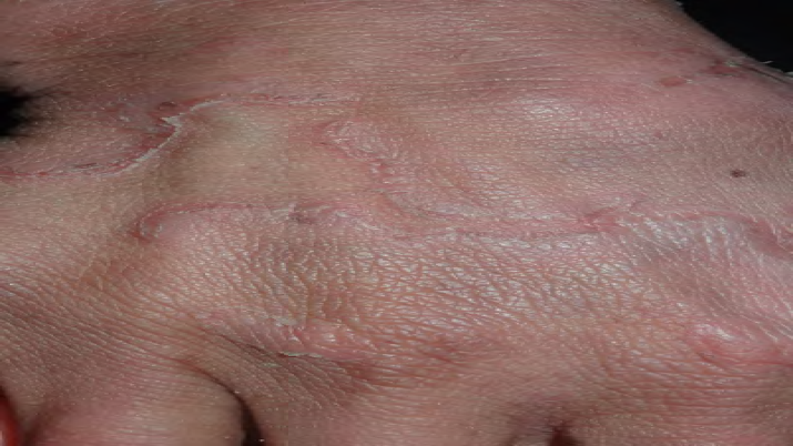 Figure 1:Typical ichthyosis linearis circumflexa lesion on the dorsum of the
right hand.
Figure 1:Typical ichthyosis linearis circumflexa lesion on the dorsum of the
right hand.
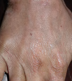 Figure 2:Complete resolution of ichthyosis linearis circumflexa 6 months
after initiation of acitretin.
Figure 2:Complete resolution of ichthyosis linearis circumflexa 6 months
after initiation of acitretin.
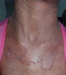 Figure 3:At presentation. Ichthyosis linearis circumflexa on the upper chest
and generalized lentiginosis.
Figure 3:At presentation. Ichthyosis linearis circumflexa on the upper chest
and generalized lentiginosis.
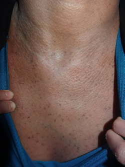 Figure 4:Six months after acitretin initiation. Complete resolution of the
ichthyosis linearis circumflexa lesions. Note the lentigines.
Figure 4:Six months after acitretin initiation. Complete resolution of the
ichthyosis linearis circumflexa lesions. Note the lentigines.
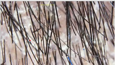 Figure 5:Trichoscopic image of the scalp hair showing trichorrhexis
invaginata (red arrow) and a golf-tee hair (blue arrow).
Figure 5:Trichoscopic image of the scalp hair showing trichorrhexis
invaginata (red arrow) and a golf-tee hair (blue arrow).
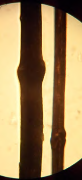 Figure 6:Case 2. Light microscopic image of a bamboo hair.
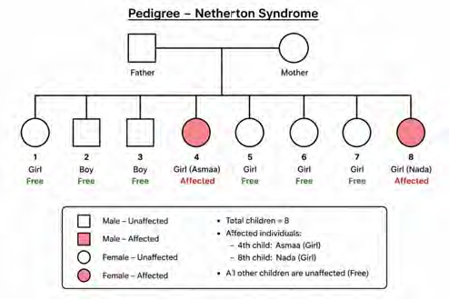 Figure 7:Pedigree of the family.
Case Report
Netherton Syndrome in Two Sisters, One with Generalized Lentiginosis: A Case Report from Yemen
Alshami MA1*, Alshami AM2, Alshami HM1 and Lutf RM1
1. Department of Dermatology, Faculty of Medicine and Medical Sciences,
Sana’a University, Sana’a, Yemen
2 Department of Conservative Dentistry, Faculty of Dentistry, Sana’a University, Sana’a, Yemen
2 Department of Conservative Dentistry, Faculty of Dentistry, Sana’a University, Sana’a, Yemen
*Address for Correspondence:Mohammad Ali Alshami, Department of Dermatology, Faculty of
Medicine and Medical Sciences, Sana’a University, Sana’a 1064, Yemen. E-mail Id: mohammadalshami62@gmail.com
Submission: 15 June, 2026
Accepted: 23 July, 2026
Published: 26 July, 2026
Copyright: © 2026 Alshami MA, et al. This is an open access
article distributed under the Creative Commons Attribution License,
which permits unrestricted use, distribution, and reproduction in any
medium, provided the original work is properly cited.
Keywords:Netherton Syndrome; Congenital Ichthyosiform
Erythroderma; Trichorrhexis invaginata; Ichthyosis Linearis Circumflexa;
Hyper-Ige
Abstract
Netherton syndrome is a rare, multisystem, autosomal recessive
genodermatosis characterized by the triad of atopic manifestations,
congenital ichthyosiform erythroderma, and trichorrhexis invaginata
(TI). We describe two sisters, aged 9 and 21 years, who presented to
our dermatology outpatient clinic with short, brittle hair, ichthyosis,
hypereosinophilia, and elevated immunoglobulin E (IgE) levels. The
older sister also exhibited generalized lentiginosis. Dermoscopy of the
scalp and eyebrow hairs demonstrated the characteristic features
of TI, which were confirmed by light microscopic examination of
plucked hairs. The clinical diagnosis of Netherton syndrome was based
on the characteristic cutaneous features, hair shaft abnormalities,
hypereosinophilia, and elevated serum IgE levels.
Introduction
Netherton syndrome (NS) is an autosomal recessive
genodermatosis caused by pathogenic variants in SPINK5.[1] It
is characterized by the triad of atopic manifestations, congenital
ichthyosiform erythroderma, and trichorrhexis invaginata (TI),
with an estimated incidence of 1 in 100,000–200,000 live births.[2]
Ichthyosis linearis circumflexa (ILC), a hallmark feature of NS, may
develop later in childhood or adulthood.[3] TI may be difficult to
detect because only a subset of hairs, most commonly the eyebrows,
is affected.[4] We report two sisters who presented with ichthyosis,
short, brittle hair, food allergies, hypereosinophilia, and elevated
serum immunoglobulin E (IgE) levels and were clinically diagnosed
with NS. Notably, the older sister also presented with the rare finding
of generalized lentiginosis.[5]
Case Report
Two sisters, aged 9 and 21 years, presented with a generalized
rash, pruritus, xerosis, and short, brittle hair, all of which had been
present since birth (Figures 1–6). They were the fourth and eighth
of eight siblings. Both were born to first-degree consanguineous
parents and presented with erythroderma at birth (Figure 7). They
also had recurrent cutaneous infections and allergic rhinitis. Since
birth, both patients had dry, cracked skin, which had previously been
regarded as mild ichthyosis and treated with emollients. Physical
examination of both sisters revealed extensive xerosis and short,
brittle hair. Peripheral eosinophilia was present in both patients, with
eosinophil counts of 1184 cells/μL (16%) and 1258 cells/μL (17%),
respectively. Total serum IgE levels were elevated at 2,500 and 1,160
IU/mL, respectively (normal value, <200 IU/mL). Trichoscopy and
light microscopic examination of the scalp and eyebrow hairs in both
sisters demonstrated the typical features of TI (Figures 5) (Figure 6).
The diagnosis of NS was established based on the presence of allergic
disease (i.e., atopic dermatitis, atopic diathesis, food allergies, hay
fever, a history of anaphylaxis, elevated IgE levels, and eosinophilia),
together with one or more of the following criteria:
1. Typical skin lesions – Scaling erythroderma, ILC. (present)
2. Typical hair findings – TI, “golf-tee,” and “matchstick” hairs. (present)
3. Family history – History of NS in a sibling .(present)
4. Genetic testing – Identification of biallelic pathogenic germline SPINK5 variants by DNA sequencing confirms the diagnosis in up to 75% of cases meeting the clinical diagnostic criteria. (Not performed because of the lack of testing facilities).[6]
Taken together, the erythroderma, ILC, elevated serum IgE levels, and atopic features supported the clinical diagnosis of NS. Both patients were initially treated with topical corticosteroids and emollients (Figures 1) (Figure 3). Given the reported safety and efficacy of oral acitretin for NS and other disorders of keratinization, we elected to initiate oral acitretin therapy at a dose of 0.62–1 mg/kg.[7] The treatment was well tolerated, with liver enzyme and lipid profile results remaining within normal limits. Mild cheilitis was observed but was successfully managed with a moisturizer. Overall, treatment markedly improved ILC, whereas TI remained unchanged. Genetic testing was not performed because of the lack of testing facilities, which represents a limitation of the present report. At the 6-month follow-up visit, both sisters showed near-complete resolution of their skin lesions (ILC), whereas TI showed no improvement (Figures 2) (Figure 4). Both patients remain under close dermatologic follow-up
2. Typical hair findings – TI, “golf-tee,” and “matchstick” hairs. (present)
3. Family history – History of NS in a sibling .(present)
4. Genetic testing – Identification of biallelic pathogenic germline SPINK5 variants by DNA sequencing confirms the diagnosis in up to 75% of cases meeting the clinical diagnostic criteria. (Not performed because of the lack of testing facilities).[6]
Taken together, the erythroderma, ILC, elevated serum IgE levels, and atopic features supported the clinical diagnosis of NS. Both patients were initially treated with topical corticosteroids and emollients (Figures 1) (Figure 3). Given the reported safety and efficacy of oral acitretin for NS and other disorders of keratinization, we elected to initiate oral acitretin therapy at a dose of 0.62–1 mg/kg.[7] The treatment was well tolerated, with liver enzyme and lipid profile results remaining within normal limits. Mild cheilitis was observed but was successfully managed with a moisturizer. Overall, treatment markedly improved ILC, whereas TI remained unchanged. Genetic testing was not performed because of the lack of testing facilities, which represents a limitation of the present report. At the 6-month follow-up visit, both sisters showed near-complete resolution of their skin lesions (ILC), whereas TI showed no improvement (Figures 2) (Figure 4). Both patients remain under close dermatologic follow-up
Discussion
Trichoscopy of the eyebrow and scalp hairs in both sisters
demonstrated the typical features of NS, namely bamboo, golf-tee,
and matchstick hairs, along with less common findings such as pili
torti and trichorrhexis nodosa (Figures 5) (Figure 6).[8]
Both sisters presented with features typically reported in NS.
The older sister additionally presented with generalized lentiginosis,
which has previously been reported only once by Xu et al. in a Chinese
patient with NS. [5,9] This phenotypic difference raises questions
regarding the genotype–phenotype correlation in NS, considering
that both sisters presumably inherited the same SPINK5 variant.
[10] However, intrafamilial and interfamilial phenotypic variation
has previously been reported in patients with NS. Furthermore,
the clinical presentation in the present case is consistent with the
findings reported by Xu et al. Generalized lentiginosis in NS has also
been reported by Moutran et al. following prolonged narrowband
ultraviolet B phototherapy.[9]
References
Citation
Alshami MA, Alshami AM, Alshami HM, Lutf RM. Netherton Syndrome in Two Sisters, One with Generalized Lentiginosis: A Case Report from Yemen. J Clin Investigat Dermatol. 2026;14(1): 1