

Journal of Clinical and Investigative Dermatology
Download PDF
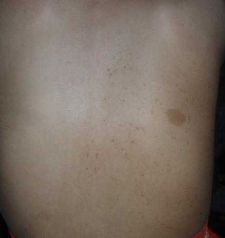 Figure 1:Unilateral café-au-lait macule associated with localized freckling on
the right side of the back, demonstrating a mosaic distribution pattern
Figure 1:Unilateral café-au-lait macule associated with localized freckling on
the right side of the back, demonstrating a mosaic distribution pattern
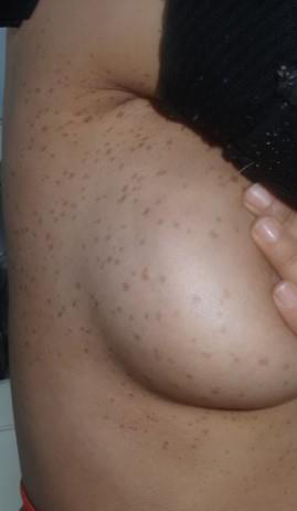 Figure 2:Lentigines involving the right axilla, breast, and abdomen.
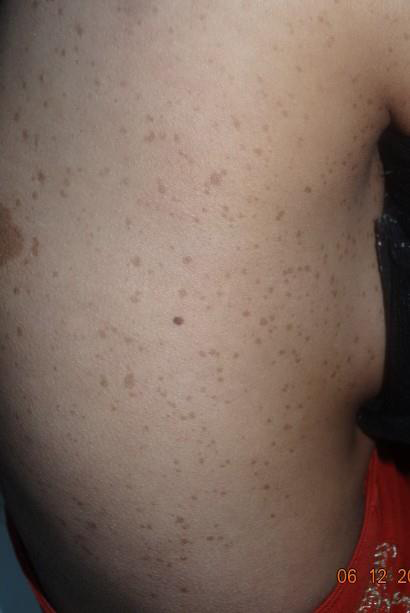 Figure 3:Right lateral view showing the large café-au-lait macule and
lentigines covering the right side of the trunk.
Figure 3:Right lateral view showing the large café-au-lait macule and
lentigines covering the right side of the trunk.
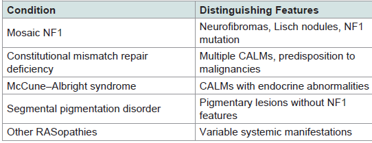 Table 1:Additional differential diagnoses.
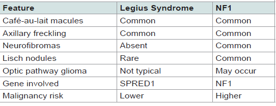 Table 2:Clinical features distinguishing Legius syndrome from NF1.
Case Report
Clinically Suspected Mosaic Legius Syndrome in a Yemeni Adolescent: A Rare Case Report
Alshami MA1*, Alshami AM2, Alshami HM1 and Lutf RM1
1Department of Dermatology, Faculty of Medicine and Medical Sciences,
Sana’a University, Yemen
2Department of Conservative Dentistry, Faculty of Dentistry, Sana’a University, Yemen
2Department of Conservative Dentistry, Faculty of Dentistry, Sana’a University, Yemen
*Address for Correspondence:Mohammad Ali Alshami, Department of Dermatology, Faculty of
Medicine and Medical Sciences, Sana’a University, Sana’a 1064, Yemen. E-mail Id: mohammadalshami62@gmail.com
Submission: 13 April, 2026
Accepted: 23 June, 2026
Published: 26 June, 2026
Copyright: © 2026 Alshami MA, et al. This is an open access
article distributed under the Creative Commons Attribution License,
which permits unrestricted use, distribution, and reproduction in any
medium, provided the original work is properly cited.
Keywords: Legius Syndrome; Mosaicism; Café-Au-Lait Macules;
Neurofibromatosis Type 1; Partial Unilateral Lentiginosis
Abstract
Legius syndrome, also known as neurofibromatosis type 1 (NF1)-
like syndrome, is an autosomal dominant condition characterized by
café-au-lait macules (CALMs) and freckling in the axilla or other sites,
without neurofibromas. Herein, we present a 14-year-old girl with a
two-year history of four CALMs on the forehead, left temple, left arm,
and right side of the back. Cutaneous examination revealed lentigines
on the axilla, chest, and right side of the back. Mosaic NF1 was
considered due to unilateral CALMs and axillary freckling; however,
neurofibromas were absent. Based solely on typical clinical findings,
mosaic Legius syndrome was diagnosed.
Introduction
Legius syndrome, also known as neurofibromatosis type 1 (NF1)-
like syndrome, is an autosomal dominant disorder first described by
Brems et al. in 2007 [1] It is characterized by café-au-lait macules
(CALMs) and axillary or intertriginous freckling in the absence of the
neurofibromas typically observed in NF1 [1] Additional dysmorphic
features may include hypertelorism, macrocephaly, lipomas, mild
learning difficulties, and attention deficits [2,3]. The disorder is
caused by pathogenic variants in the SPRED1 gene, which encodes
Sprouty-related EVH1 domain–containing protein 1 (SPRED1), a
negative regulator of the RAS-MAPK signaling pathway involved in
cellular growth and development [3]. Herein, we describe a clinically
diagnosed case of mosaic Legius syndrome in an adolescent girl.
Case report
A 14-year-old girl presented with a two-year history of multiple
CALMs on the forehead, left temple, left arm, and right side of the
back (Figures 1–3). Cutaneous examination revealed freckling over
the axilla, chest, and right side of the back. No neurofibromas, learning
disabilities or developmental delays were identified. Ophthalmologic
examination showed no evidence of Lisch nodules, and orthopedic
evaluation revealed no osseous abnormalities characteristic of NF.
Partial unilateral lentiginosis (PUL) was initially considered owing to
the segmental distribution of the lesions on the right side of the trunk;
however, the presence of four CALMs and ipsilateral axillary freckling
complicated the diagnosis. Literature search revealed similar cases of
PUL with CALMs and additional axillary freckling, while a few reports
described co-occurrence with Lisch nodules. The concomitance of
PUL, axillary freckling, and CALMs in the present case raised the
possibility of mosaic neurofibromatosis; however, the absence of
neurofibromas rendered this diagnosis less likely and instead favored
a diagnosis of Legius syndrome [4-6] Other differential diagnoses are
listed in (Table 1). No family members exhibited similar cutaneous
findings. Based on the clinical presentation, a diagnosis of mosaic
Legius syndrome was made; however, molecular confirmation was
not available. Because the patient was only concerned about the
cosmetic appearance of the CALMs, treatment was initiated with a
Q-switched Nd:YAG laser, followed by topical adapalene gel and kojic
acid. Four months after two treatment sessions, patient satisfaction
and objective clinical improvement were observed.
This case underscores the diagnostic complexity of mosaic pigmentary disorders, particularly when clinical features overlap with those of mosaic NF1(Table 1).
This case underscores the diagnostic complexity of mosaic pigmentary disorders, particularly when clinical features overlap with those of mosaic NF1(Table 1).
Discussion
Mosaic Legius syndrome closely resembles mosaic NF1 but is
distinguished by the absence of neurofibromas and generally exhibits
a milder clinical phenotype [7]. Key clinical features that aid in
differentiating Legius syndrome from NF1 are summarized in Table
2. However, recent reports of Lisch nodules in two siblings with Legius
syndrome suggest that the phenotypic distinction between Legius
syndrome and NF1 may be less absolute than previously recognized
[8] Accurate diagnosis is therefore essential for differentiating
Legius syndrome from other disorders with similar features, such
as NF1, as it can prevent unnecessary malignancy surveillance and
alleviate familial anxiety regarding NF1-related complications [9]
Nevertheless, reports of SPRED1-associated leukemia suggest that
some degree of clinical vigilance may still be warranted [10].
Although genetic testing for SPRED1 variants could have confirmed our diagnosis, such testing was not available. Nonetheless, this clinically diagnosed case highlights a rare mosaic presentation of Legius syndrome and expands the limited dermatologic literature from resource-constrained settings. Regarding the treatment of PUL and CALMs, available evidence supports the efficacy and safety of Q-switched Nd: YAG laser. Accordingly, this treatment was initiated because the patient was concerned only about the cosmetic burden associated with CALMs.
Although genetic testing for SPRED1 variants could have confirmed our diagnosis, such testing was not available. Nonetheless, this clinically diagnosed case highlights a rare mosaic presentation of Legius syndrome and expands the limited dermatologic literature from resource-constrained settings. Regarding the treatment of PUL and CALMs, available evidence supports the efficacy and safety of Q-switched Nd: YAG laser. Accordingly, this treatment was initiated because the patient was concerned only about the cosmetic burden associated with CALMs.
References
Citation
Alshami MA, Alshami AM, Alshami HM, Lutf RM. Clinically Suspected Mosaic Legius Syndrome in a Yemeni Adolescent: A Rare Case Report. J Clin Investigat Dermatol. 2026;14(1): 1