

Journal of Chemistry and Applications
Download PDF
The DPPH assay was performed in triplicate (n = 3), and the results are expressed as mean ± standard deviation (SD).
The percentage of DPPH radical scavenging activity was calculated using the following equation:
DPPH scavenging activity (%) = [(A₀ − Aₜ)/A₀] × 100
where A₀ represents the absorbance of the control and Aₜ represents the absorbance of the test sample.
In addition to evaluating antioxidant activity, the synthesized AuNPs were further investigated for their antibacterial and anticancer activities to explore their potential biomedical applications.
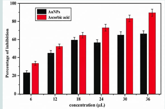 Figure 1:UV–Visible absorption spectra of AuNPs synthesized using (a) different concentrations of CSFE, (b) different concentrations of HAuCl₄, and (c) different autoclave reaction times.
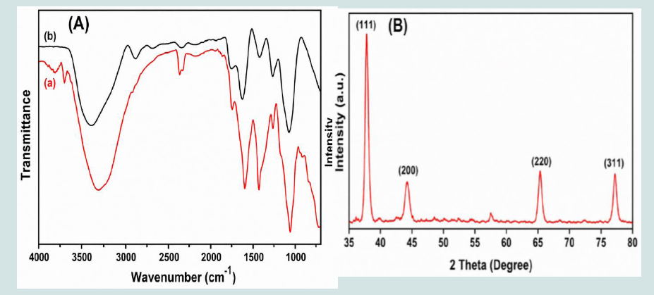 Figure 2:(A) FTIR spectra of (a) CSFE and (b) CSFE-capped gold nanoparticles (AuNPs). (B) X-ray diffraction (XRD) pattern of the synthesized AuNPs.
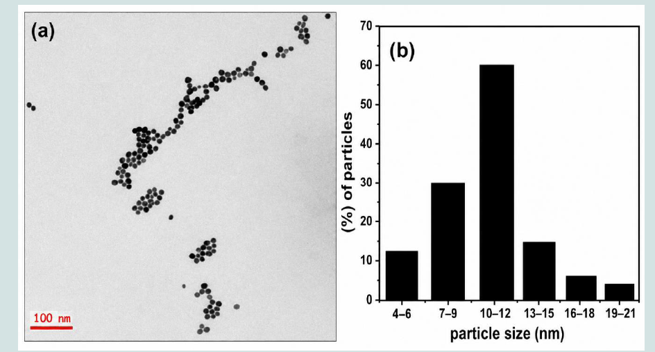 Figure 3:(a) TEM micrograph of the synthesized CSFE AuNPs and (b) particle size distribution histogram of the synthesized AuNPs.
Figure 4:DPPH free radical scavenging activity of biosynthesized CSFE AuNPs at different concentrations compared with ascorbic acid. Data are presented as mean ± SD (n = 3). Error bars represent the standard deviation.
Figure 3:(a) TEM micrograph of the synthesized CSFE AuNPs and (b) particle size distribution histogram of the synthesized AuNPs.
Figure 4:DPPH free radical scavenging activity of biosynthesized CSFE AuNPs at different concentrations compared with ascorbic acid. Data are presented as mean ± SD (n = 3). Error bars represent the standard deviation.
The DPPH assay was performed in triplicate (n = 3), and the results are presented as mean ± standard deviation (SD). Error bars representing the standard deviation are included in Figure 4 to demonstrate the reproducibility and reliability of the experimental results.
As shown in Figure 4, the free radical scavenging activity of the synthesized AuNPs increased with increasing nanoparticle concentration, indicating concentration-dependent antioxidant activity. The percentage inhibition values observed for the AuNPs were 23.2%, 45.6%, 59.1%, 56.3%, 63.1%, and 65.25% at concentrations of 6, 12, 18, 24, 30, and 36 μL, respectively. These results demonstrate that the biosynthesized AuNPs possess significant antioxidant activity, which can be attributed to the phytochemical constituents adsorbed on the nanoparticle surface. The antioxidant performance of the synthesized AuNPs was comparable to that of the standard antioxidant, ascorbic acid, particularly at higher concentrations.
Research Article
Sustainable Fabrication of Gold Nanoparticles Using Catunaregam spinosa Fruit Extract for Antioxidant Applications
Banu R1* and Mohan KC2
1Department of Chemistry, Dr. BRR GDC(A) Jadcherla,
Mahabubnagar Telangana, India.
2Department of Chemistry, MALD Government Degree College Gadwal Telangana, India.
2Department of Chemistry, MALD Government Degree College Gadwal Telangana, India.
*Address for Correspondence:Ruqya Banu, Department of Chemistry, Dr. BRR GDC(A) Jadcherla, Mahabubnagar Telangana, India. EMail Id:ruqyabrr@gmail.com
Submission: 20 June, 2026
Accepted: 28 July, 2026
Published: 30 July, 2026
Copyright: © 2026 Banu R, et al. This is an open access article
distributed under the Creative Commons Attribution License, which
permits unrestricted use, distribution, and reproduction in any medium,
provided the original work is properly cited.
Keywords: Gold Nanoparticles; Catunaregam Spinosa; Green
Synthesis; Antioxidant Activity; DPPH Assay.
Abstract
The green synthesis of gold nanoparticles (AuNPs) has emerged as
a promising and sustainable approach in nanotechnology owing to its
environmental friendliness, cost-effectiveness, and simplicity. In the present
study, a rapid and eco-friendly method was developed for the synthesis
of AuNPs using Catunaregam spinosa fruit extract as a natural reducing,
capping, and stabilizing agent. The biosynthesized AuNPs exhibited an
average particle size of 10 ± 2 nm. Comprehensive characterization of the
synthesized nanoparticles was carried out using UV–Visible spectroscopy,
Fourier transform infrared (FTIR) spectroscopy, X-ray diffraction (XRD),
and transmission electron microscopy (TEM). The formation of AuNPs
was confirmed by the characteristic surface plasmon resonance (SPR)
absorption band in the UV–Visible spectrum, while XRD analysis revealed
their crystalline nature with a face-centered cubic (FCC) structure. TEM
analysis demonstrated that the nanoparticles were predominantly
spherical and well dispersed. The antioxidant potential of the synthesized
AuNPs was evaluated using the 2,2-diphenyl-1-picrylhydrazyl (DPPH) free
radical scavenging assay, which demonstrated significant concentrationdependent
antioxidant activity. These findings indicate that Catunaregam
spinosa fruit extract-mediated AuNPs represent an environmentally
sustainable nanomaterial with promising potential for biomedical and
antioxidant applications.
Introduction
Nanoscience and nanotechnology have emerged as rapidly
advancing multidisciplinary fields that integrate principles from
chemistry, biology, physics, and engineering to design, synthesize,
and manipulate materials at the nanoscale. Owing to their
unique physicochemical properties, nanomaterials have attracted
considerable attention in both academic research and industrial
applications. Among them, metallic nanoparticles (MNPs) have
gained particular interest because their optical, electrical, catalytic,
and biological properties are strongly dependent on particle size and
morphology, distinguishing them from their bulk counterparts [1].
The unique physicochemical properties of MNPs, including their high surface-to-volume ratio, enhanced surface reactivity, and tunable optical characteristics, have enabled their application in diverse fields such as antimicrobial agents, biosensors, catalysis, drug delivery, water purification, and biomedical imaging [2]. Among the various noble metal nanoparticles, gold nanoparticles (AuNPs) have attracted significant attention owing to their excellent biocompatibility, chemical stability, and remarkable optical properties. These characteristics make AuNPs highly suitable for applications in catalysis, sensing, photonics, biomedical diagnostics, targeted drug delivery, imaging, and antimicrobial therapy.
The unique physicochemical properties of MNPs, including their high surface-to-volume ratio, enhanced surface reactivity, and tunable optical characteristics, have enabled their application in diverse fields such as antimicrobial agents, biosensors, catalysis, drug delivery, water purification, and biomedical imaging [2]. Among the various noble metal nanoparticles, gold nanoparticles (AuNPs) have attracted significant attention owing to their excellent biocompatibility, chemical stability, and remarkable optical properties. These characteristics make AuNPs highly suitable for applications in catalysis, sensing, photonics, biomedical diagnostics, targeted drug delivery, imaging, and antimicrobial therapy.
Several methods have been developed for the synthesis of
AuNPs, including chemical, physical, and biological approaches.
Conventional chemical methods generally involve the reduction
of gold salts using reducing agents such as sodium borohydride,
hydrazine, citrate, and other synthetic chemicals [3]. Although these
methods efficiently produce nanoparticles with well-controlled size
and morphology, they often require hazardous reagents, consume
substantial energy, and generate toxic by-products, thereby limiting
their environmental sustainability and biomedical applicability.
In recent years, green synthesis has emerged as an environmentally
benign and sustainable alternative for nanoparticle production.
Biological resources such as microorganisms, fungi, algae, and plant
extracts have been extensively explored as eco-friendly reducing and
stabilizing agents for the synthesis of metal nanoparticles [4]. Among
these biological resources, plant-mediated synthesis is particularly
advantageous because plant extracts are rich in naturally occurring
phytochemicals that simultaneously function as reducing, capping,
and stabilizing agents. This eliminates the need for toxic chemicals
and provides a simple, rapid, cost-effective, and environmentally
friendly route for nanoparticle synthesis.
More recently, biomass-derived materials have attracted
considerable interest as renewable resources for sustainable
nanotechnology. Biomass materials, including medicinal plants,
agricultural residues, natural polymers, and other renewable
biological resources, contain abundant bioactive compounds such
as polyphenols, flavonoids, alkaloids, proteins, carbohydrates,
and organic acids, which facilitate the reduction of metal ions and
stabilize the resulting nanoparticles. Compared with conventional
chemical synthesis, biomass-assisted approaches offer several
advantages, including reduced environmental impact, lower
production costs, enhanced biocompatibility, improved nanoparticle
stability, and greater sustainability. Consequently, biomass-mediated
nanomaterials have found promising applications in catalysis,
environmental remediation, sensing, energy storage, antimicrobial
therapy, antioxidant activity, and drug delivery [5-9].
Catunaregam spinosa is a medicinal plant widely used in
traditional Ayurvedic medicine because of its diverse therapeutic
properties. The fruits of C. spinosa are rich in phytochemicals,
including flavonoids, alkaloids, phenolic compounds, coumarins,
steroids, carbohydrates, and other bioactive constituents. These
naturally occurring compounds not only contribute to the medicinal
value of the fruit but also serve as effective reducing, capping, and
stabilizing agents during the green synthesis of metal nanoparticles
[10].
Although numerous plant extracts have been employed for
the green synthesis of gold nanoparticles, the use of Catunaregam
spinosa fruit extract as a renewable biomass source remains largely
unexplored. Therefore, the novelty of the present study lies in the
rapid and eco-friendly synthesis of stable AuNPs using Catunaregam
spinosa fruit extract without the use of hazardous chemical reducing
agents. In addition, the structural, morphological, catalytic, and
antioxidant properties of the synthesized AuNPs were systematically
investigated to evaluate their potential as environmentally sustainable
nanomaterials for biomedical and environmental applications.
In this study, AuNPs were successfully synthesized
using Catunaregam spinosa fruit extract through a rapid and
environmentally friendly green synthesis approach. The synthesized
nanoparticles were comprehensively characterized using UV–Visible
spectroscopy, Fourier transform infrared (FTIR) spectroscopy, X-ray
diffraction (XRD), energy-dispersive X-ray spectroscopy (EDX),
and transmission electron microscopy (TEM) [11]. Furthermore,
the catalytic performance of the synthesized AuNPs was evaluated
through the reduction of methylene blue dye, while their antioxidant
activity was assessed using the 2,2-diphenyl-1-picrylhydrazyl
(DPPH) free radical scavenging assay. The results demonstrate
that Catunaregam spinosa fruit extract-mediated AuNPs are
environmentally sustainable nanomaterials with promising potential
for catalytic, antioxidant, and future biomedical applications.
Materials and Methods
Materials:
Hydrogen tetrachloroaurate trihydrate (HAuCl₄·3H₂O) was
procured from Sigma-Aldrich (India). All other chemicals and
reagents used in this study were of analytical grade and purchased
from Merck (India). Double-distilled water was used throughout
all experiments. Before use, all glassware was thoroughly cleaned
with freshly prepared aqua regia, followed by repeated rinsing with
double-distilled water to remove any residual contaminants.
Preparation of Catunaregam spinosa Fruit Extract (CSFE):
Fresh fruits of Catunaregam spinosa were collected from the
Mannanur forest region, Mahabubnagar, Telangana, India. The fruits
were thoroughly washed several times with double-distilled water
to remove dust and other surface impurities. Approximately 6 g of
fresh fruit material was crushed and mixed with 60 mL of doubledistilled
water. The mixture was heated at 64–70 °C for 50 min and
then allowed to cool to room temperature. The resulting extract
was filtered through Whatman No. 1 filter paper, and the clear
filtrate was collected and stored for the subsequent synthesis of gold
nanoparticles.
Green Synthesis of Gold Nanoparticles:
Gold nanoparticles (AuNPs) were synthesized using a green,
plant-mediated approach. Briefly, 5 mL of CSFE was mixed with 4
mL of 1 mM aqueous HAuCl₄ solution. The reaction mixture was
transferred to an autoclave and maintained at 120 °C under a pressure
of 15 psi for 20 min. Upon completion of the reaction, the solution
developed a characteristic ruby-red color, indicating the reduction of
Au³ ions to metallic gold (Au⁰) and the successful formation of gold
nanoparticles [12].
Characterization of Gold Nanoparticles:
The optical properties of the synthesized AuNPs were analyzed
using a UV–Visible spectrophotometer (UV-3600, Shimadzu, Japan).
Fourier transform infrared (FTIR) spectra were recorded using an
IRAffinity-1 spectrometer (Shimadzu, Japan) to identify the functional
groups responsible for the reduction, capping, and stabilization of the
nanoparticles [13-14]. The crystalline structure of the synthesized
AuNPs was investigated by X-ray diffraction (XRD) using a Rigaku
Miniflex diffractometer equipped with Cu-Kα radiation (λ = 1.5406
Å). The morphology and particle size distribution of the nanoparticles
were examined using transmission electron microscopy (TEM; JEM-
1200EX, JEOL Ltd., Japan).
Antioxidant Activity:
The antioxidant activity of the synthesized AuNPs was evaluated
using the 2,2-diphenyl-1-picrylhydrazyl (DPPH) free radical
scavenging assay with slight modifications to a previously reported
method. Different volumes of the AuNP suspension (6, 12, 18, 24,
30, and 36 μL) were diluted with dimethyl sulfoxide (DMSO) to a
final volume of 60 μL. Subsequently, 2.56 mL of 0.17 mM DPPH
solution was added to each sample. The reaction mixtures were
vigorously mixed and incubated in the dark at room temperature for
40 min. The absorbance was measured at 517 nm using a UV–Visible
spectrophotometer. Ascorbic acid was used as the standard reference
antioxidant.The DPPH assay was performed in triplicate (n = 3), and the results are expressed as mean ± standard deviation (SD).
The percentage of DPPH radical scavenging activity was calculated using the following equation:
DPPH scavenging activity (%) = [(A₀ − Aₜ)/A₀] × 100
where A₀ represents the absorbance of the control and Aₜ represents the absorbance of the test sample.
In addition to evaluating antioxidant activity, the synthesized AuNPs were further investigated for their antibacterial and anticancer activities to explore their potential biomedical applications.
Results and Discussion
Characterization of Gold Nanoparticles:
The successful synthesis of gold nanoparticles (AuNPs) was
initially confirmed by UV–Visible spectroscopy through the
appearance of the characteristic surface plasmon resonance (SPR)
absorption band. The synthesized AuNPs exhibited a distinct SPR
peak in the wavelength range of 520–530 nm, which is characteristic
of spherical gold nanoparticles. The influence of CSFE concentration
(0.1–0.8%) on nanoparticle synthesis was investigated, and the
corresponding UV–Visible spectra are presented in Figure 1a. As
the concentration of CSFE increased, the intensity of the SPR band
gradually increased, indicating enhanced reduction of Au³⁻ ions and
increased formation of AuNPs. This observation suggests that the
phytochemical constituents present in the fruit extract play a vital
role in the reduction and stabilization of gold nanoparticles [15].
The effect of the gold precursor concentration on nanoparticle
synthesis was also investigated using different concentrations of
HAuCl₄, as shown in Figure 1b. An increase in HAuCl₄ concentration
resulted in a corresponding increase in the intensity of the SPR
band, indicating a higher yield of AuNPs under increased precursor
concentrations [16]. The influence of autoclave reaction time on
nanoparticle formation was evaluated and is presented in Figure
1c. As the reaction time increased from 0.5 to 20 min, the SPR
peak intensity progressively increased, reflecting the formation of
a greater number of AuNPs. However, extending the reaction time
beyond 20 min did not produce any significant changes in either the
intensity or the shape of the SPR band, indicating that the reduction
reaction had reached completion. These results demonstrate that
the concentrations of both the plant extract and the gold precursor,
together with the reaction time, play crucial roles in determining the
efficiency of AuNP synthesis [17-18].
The Fourier transform infrared (FTIR) spectra of the CSFE and the biosynthesized AuNPs were compared to identify the functional groups involved in the reduction, capping, and stabilization of the nanoparticles (Figure 2a). The observed shifts in peak positions and changes in peak intensities after nanoparticle synthesis confirm the interaction of phytochemical constituents with the surface of the AuNPs.
A broad absorption band in the region of 3400–3300 cm⁻¹ was assigned to the stretching vibration of hydroxyl (–OH) groups present in phenolic compounds, flavonoids, tannins, carbohydrates, and alcohols. Following nanoparticle synthesis, this band became broader and shifted slightly toward a lower wavenumber, indicating the involvement of hydroxyl groups in coordinating with the gold nanoparticle surface. These hydroxyl-containing phytochemicals likely donated electrons to reduce Au³⁺ ions while simultaneously stabilizing the synthesized nanoparticles.
The absorption bands observed in the 2920–2850 cm⁻¹ region correspond to the asymmetric and symmetric stretching vibrations of aliphatic –CH₂ and –CH₃ groups present in proteins and other organic constituents. Minor variations in these bands after nanoparticle synthesis suggest that aliphatic functional groups are associated with the nanoparticle surface but play only a limited role in the reduction process.
The Fourier transform infrared (FTIR) spectra of the CSFE and the biosynthesized AuNPs were compared to identify the functional groups involved in the reduction, capping, and stabilization of the nanoparticles (Figure 2a). The observed shifts in peak positions and changes in peak intensities after nanoparticle synthesis confirm the interaction of phytochemical constituents with the surface of the AuNPs.
A broad absorption band in the region of 3400–3300 cm⁻¹ was assigned to the stretching vibration of hydroxyl (–OH) groups present in phenolic compounds, flavonoids, tannins, carbohydrates, and alcohols. Following nanoparticle synthesis, this band became broader and shifted slightly toward a lower wavenumber, indicating the involvement of hydroxyl groups in coordinating with the gold nanoparticle surface. These hydroxyl-containing phytochemicals likely donated electrons to reduce Au³⁺ ions while simultaneously stabilizing the synthesized nanoparticles.
The absorption bands observed in the 2920–2850 cm⁻¹ region correspond to the asymmetric and symmetric stretching vibrations of aliphatic –CH₂ and –CH₃ groups present in proteins and other organic constituents. Minor variations in these bands after nanoparticle synthesis suggest that aliphatic functional groups are associated with the nanoparticle surface but play only a limited role in the reduction process.
The absorption band located around 1650–1600 cm⁻¹ is
attributed to the stretching vibration of carbonyl (C=O) groups of
proteins, polyphenols, and conjugated ketones and may also include
amide I vibrations. The shift of this band toward a lower wavenumber
following nanoparticle formation indicates the coordination of
carbonyl oxygen atoms with the AuNP surface, thereby contributing
to nanoparticle stabilization.
A band observed near 1540–1510 cm⁻¹ corresponds to amide II vibrations and N–H bending modes. The slight shift and reduction in intensity of this band suggest the participation of amino groups from proteins or amino acids in binding to the nanoparticle surface through nitrogen-containing functional groups.
The absorption bands in the 1450–1380 cm⁻¹ region arise from the bending vibrations of methyl groups and the symmetric stretching of carboxylate (COO⁻) ions. The observed shifts in these bands indicate the interaction of carboxyl groups from organic acids and proteins with the AuNP surface, thereby enhancing the colloidal stability of the nanoparticles.
A band observed near 1540–1510 cm⁻¹ corresponds to amide II vibrations and N–H bending modes. The slight shift and reduction in intensity of this band suggest the participation of amino groups from proteins or amino acids in binding to the nanoparticle surface through nitrogen-containing functional groups.
The absorption bands in the 1450–1380 cm⁻¹ region arise from the bending vibrations of methyl groups and the symmetric stretching of carboxylate (COO⁻) ions. The observed shifts in these bands indicate the interaction of carboxyl groups from organic acids and proteins with the AuNP surface, thereby enhancing the colloidal stability of the nanoparticles.
The absorption bands observed between 1250 and 1000 cm⁻¹ are
characteristic of C–O–C and C–O stretching vibrations associated
with alcohols, polysaccharides, glycosides, and flavonoids. A
reduction in band intensity accompanied by slight peak shifts after
nanoparticle synthesis indicates that these oxygen-containing
functional groups were adsorbed onto the nanoparticle surface and
acted as natural capping agents.
Overall, the FTIR analysis demonstrates that hydroxyl, carbonyl, carboxyl, amide, and C–O functional groups present in the phytochemicals of CSFE actively participated in the reduction of Au³⁻ ions and remained adsorbed on the surface of the synthesized AuNPs as natural reducing, capping, and stabilizing agents.
The crystalline nature of the synthesized AuNPs was investigated by X-ray diffraction (XRD), and the corresponding diffraction pattern is presented in Figure 2b. Distinct diffraction peaks were observed at
Overall, the FTIR analysis demonstrates that hydroxyl, carbonyl, carboxyl, amide, and C–O functional groups present in the phytochemicals of CSFE actively participated in the reduction of Au³⁻ ions and remained adsorbed on the surface of the synthesized AuNPs as natural reducing, capping, and stabilizing agents.
The crystalline nature of the synthesized AuNPs was investigated by X-ray diffraction (XRD), and the corresponding diffraction pattern is presented in Figure 2b. Distinct diffraction peaks were observed at
2θ values of 38.15°, 44.50°, 64.51°, and 77.42°, corresponding to the
(111), (200), (220), and (311) crystallographic planes, respectively.
These diffraction peaks are characteristic of the face-centered cubic
(FCC) crystal structure of metallic gold, confirming the successful
formation of crystalline AuNPs [19-21].
The average crystallite size of the synthesized AuNPs was estimated using the Debye–Scherrer equation and was found to be approximately 8.5 nm. This value is in good agreement with the particle size obtained from transmission electron microscopy (TEM), indicating the successful synthesis of nanoscale crystalline gold nanoparticles.
The morphology and particle size distribution of the synthesized AuNPs were further investigated using TEM. Representative TEM micrographs (Figure 3a) revealed that the nanoparticles were predominantly spherical, well dispersed, and exhibited minimal aggregation [22–23]. The average particle size was determined to be 10 ± 2 nm.
The average crystallite size of the synthesized AuNPs was estimated using the Debye–Scherrer equation and was found to be approximately 8.5 nm. This value is in good agreement with the particle size obtained from transmission electron microscopy (TEM), indicating the successful synthesis of nanoscale crystalline gold nanoparticles.
The morphology and particle size distribution of the synthesized AuNPs were further investigated using TEM. Representative TEM micrographs (Figure 3a) revealed that the nanoparticles were predominantly spherical, well dispersed, and exhibited minimal aggregation [22–23]. The average particle size was determined to be 10 ± 2 nm.
The particle size distribution histogram obtained from the
TEM measurements (Figure 3b) showed a relatively narrow
size distribution, indicating the uniform formation of AuNPs.
Furthermore, the particle size determined from TEM analysis
closely matched the crystallite size estimated from XRD, confirming
the successful synthesis of uniformly distributed nanoscale gold
nanoparticles through the green synthesis route using CSFE.
Overall, the spectroscopic and microscopic characterization results demonstrate that CSFE serves as an efficient natural reducing, capping, and stabilizing agent for the rapid green synthesis of stable, crystalline, and predominantly spherical gold nanoparticles.
Overall, the spectroscopic and microscopic characterization results demonstrate that CSFE serves as an efficient natural reducing, capping, and stabilizing agent for the rapid green synthesis of stable, crystalline, and predominantly spherical gold nanoparticles.
Antioxidant Activity:
The antioxidant activity of the biosynthesized AuNPs was
evaluated using the 2,2-diphenyl-1-picrylhydrazyl (DPPH) free
radical scavenging assay, with ascorbic acid serving as the standard
reference antioxidant. DPPH is a stable nitrogen-centered free radical
that accepts electrons or hydrogen atoms from antioxidant molecules,
resulting in its reduction and a characteristic color change from deep
purple to yellow [24-25].The DPPH assay was performed in triplicate (n = 3), and the results are presented as mean ± standard deviation (SD). Error bars representing the standard deviation are included in Figure 4 to demonstrate the reproducibility and reliability of the experimental results.
As shown in Figure 4, the free radical scavenging activity of the synthesized AuNPs increased with increasing nanoparticle concentration, indicating concentration-dependent antioxidant activity. The percentage inhibition values observed for the AuNPs were 23.2%, 45.6%, 59.1%, 56.3%, 63.1%, and 65.25% at concentrations of 6, 12, 18, 24, 30, and 36 μL, respectively. These results demonstrate that the biosynthesized AuNPs possess significant antioxidant activity, which can be attributed to the phytochemical constituents adsorbed on the nanoparticle surface. The antioxidant performance of the synthesized AuNPs was comparable to that of the standard antioxidant, ascorbic acid, particularly at higher concentrations.
Conclusion
In the present study, gold nanoparticles (AuNPs) were successfully
synthesized through a simple, rapid, and environmentally friendly
green synthesis approach using Catunaregam spinosa fruit extract.
The phytochemicals present in the extract acted as natural reducing,
capping, and stabilizing agents, enabling the formation of stable
AuNPs without the use of hazardous chemicals. Comprehensive
characterization by UV–Visible spectroscopy, FTIR, XRD, and TEM
confirmed the successful synthesis of crystalline, predominantly
spherical AuNPs with an average particle size of 10 ± 2 nm. The
synthesized nanoparticles exhibited significant concentrationdependent
antioxidant activity, demonstrating their potential as
effective free radical scavengers. Overall, the findings of this study
highlight the potential of Catunaregam spinosa-mediated AuNPs
as environmentally sustainable nanomaterials with promising
applications in catalysis, antioxidant therapy, and other biomedical
fields. Further investigations on their antibacterial, anticancer,
and in vivo biological activities are warranted to fully explore their
therapeutic potential.
References
Citation
Banu R, Mohan KC. Sustainable Fabrication of Gold Nanoparticles Using Catunaregam spinosa Fruit Extract for Antioxidant Applications. J Chem Applications 2026;6(1): 6.